 Back
The Skeletal System: Structure, Function, and Development
Study Guide - Smart Notes
 Tailored notes based on your materials, expanded with key definitions, examples, and context.
Skeletal System Overview
Introduction to the Skeletal System
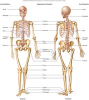
The skeletal system forms the rigid framework of the human body, providing support, protection, and facilitating movement. It is composed of bones, cartilage, ligaments, and other connective tissues, each contributing to the system's overall function and structure.
Tissues in Bone
Composition of Bone Tissue
Bones are complex organs containing several tissue types:
Bone connective tissue – the primary tissue, providing structure and strength.
Nervous tissue – present for sensation and regulation.
Blood connective tissue – found within marrow spaces and blood vessels.
Cartilage – especially in articular surfaces for joint movement.
Epithelial tissue – lines blood vessels within bone.
Functions of Bones
Major Roles of the Skeletal System
Support: Provides a hard framework for the body.
Movement: Skeletal muscles use bones as levers to produce movement.
Protection: Shields vital organs (e.g., skull protects the brain).
Mineral Storage: Reservoir for calcium and phosphate.
Blood Cell Formation: Red marrow produces blood cells (hematopoiesis).
Energy Metabolism: Osteoblasts secrete osteocalcin, influencing energy metabolism.
Bone Tissue Structure
Organic and Inorganic Components
Organic components: Cells, collagen fibers, and ground substance; provide flexibility and tensile strength (35% of bone mass).
Inorganic components: Mineral salts (mainly hydroxyapatite); provide hardness and resistance to compression (65% of bone mass).
Extracellular Matrix
The unique composition of the bone matrix gives bone its exceptional properties, balancing flexibility and strength with hardness and durability.
Bone Cells
Types of Bone Cells
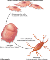
Osteoprogenitor cells: Stem cells that differentiate into osteoblasts.
Osteoblasts: Actively produce and secrete bone matrix (osteoid).
Osteocytes: Mature bone cells that maintain the bone matrix.
Osteoclasts
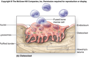
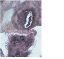
Osteoclasts are large, multinucleated cells responsible for bone resorption. They secrete hydrochloric acid and lysosomal enzymes to break down bone tissue and are derived from white blood cell lineages.
Classification of Bones
Types of Bones by Shape
Long bones: Longer than wide; consist of a shaft (diaphysis) and two ends (epiphyses). Example: humerus.
Short bones: Cube-shaped. Example: talus.
Flat bones: Thin, flattened, and usually curved. Example: sternum.
Irregular bones: Complex shapes that do not fit other categories. Example: vertebra.
Gross Anatomy of Bones
Structure of a Typical Long Bone
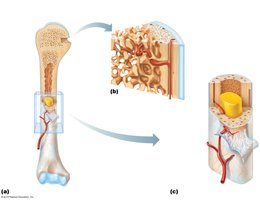
Diaphysis: The shaft of the bone.
Epiphyses: The ends of the bone.
Medullary cavity: Central cavity containing yellow marrow (fat storage).
Compact bone: Dense outer layer.
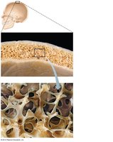
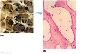
Spongy bone: Internal network of trabeculae filled with marrow.
Periosteum: Outer membrane covering the bone.
Endosteum: Membrane lining the medullary cavity and trabeculae.
Structure of Short, Irregular, and Flat Bones
These bones contain bone marrow but lack a true marrow cavity. The internal spongy bone of flat bones is called diploë.
Bone Markings
Categories of Bone Markings
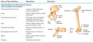
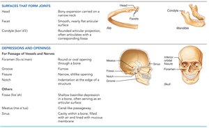
Bone markings reflect stresses and serve as sites for muscle attachment, joint formation, and passage of nerves and blood vessels.
Name of Bone Marking | Description | Illustration |
|---|---|---|
Projections (e.g., tuberosity, crest, trochanter, line, tubercle, epicondyle, spine, process) | Sites of muscle and ligament attachment | See illustration |
Name of Bone Marking | Description | Illustration |
|---|---|---|
Surfaces (e.g., head, facet, condyle, ramus) | Form joints | See illustration |
Depressions and Openings (e.g., foramen, groove, fissure, notch, fossa, meatus, sinus) | Passage of vessels and nerves | See illustration |
Microscopic Structure of Bone
Compact Bone
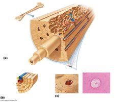
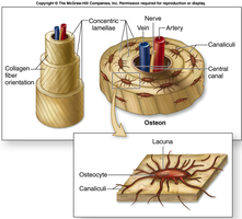
Compact bone contains passageways for blood vessels, lymph vessels, and nerves. The basic structural unit is the osteon (Haversian system), which resembles rings of a tree in cross-section.
Osteon Structure
Lamellae: Concentric rings of bone matrix.
Central canal: Contains blood vessels and nerves.
Perforating (Volkmann's) canals: Connect central canals.
Canaliculi: Tiny canals connecting osteocytes in their lacunae.
Spongy Bone
Spongy bone is less complex, consisting of trabeculae with layers of lamellae and osteocytes. It does not contain osteons.
Bone Development (Ossification)
Intramembranous Ossification
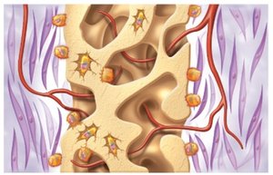
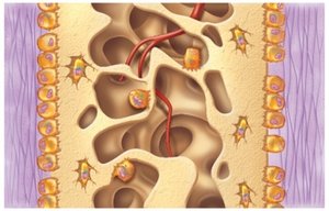
Membranous bones form directly from mesenchyme through intramembranous ossification. This process is typical for flat bones of the skull and clavicle.
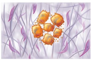
Mesenchymal cells differentiate into osteoblasts, forming ossification centers.
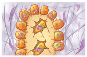
Osteoblasts secrete osteoid, which calcifies and traps some cells as osteocytes.
Woven bone and periosteum form, followed by remodeling into compact and spongy bone.
Endochondral Ossification
Most bones develop from hyaline cartilage models through endochondral ossification. This process begins in the embryo and continues into early adulthood.
Bone collar forms around the diaphysis of the cartilage model.
Cartilage in the center calcifies and develops cavities.
Periosteal bud invades, forming spongy bone.
Diaphysis elongates, medullary cavity forms, and secondary ossification centers appear in epiphyses.
Epiphyses ossify; hyaline cartilage remains only in epiphyseal plates and articular cartilage.
Growth of Bones
Epiphyseal Plate Anatomy and Growth
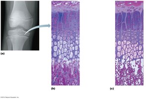
The epiphyseal plate is responsible for longitudinal bone growth during childhood and adolescence. It consists of zones where cartilage cells divide, enlarge, calcify, and are replaced by bone tissue.
Postnatal Bone Growth
Bones lengthen by growth of the epiphyseal plates, maintaining constant thickness as cartilage is replaced by bone.
As adolescence ends, chondroblasts divide less, plates thin, and eventually diaphysis and epiphysis fuse, ending growth.
Bones also widen by appositional growth (addition of bone tissue to the surface).
Hormonal Regulation
Growth hormone: Stimulates epiphyseal plate activity.
Thyroid hormone: Ensures proper skeletal proportions.
Sex hormones: Promote growth and later induce closure of epiphyseal plates.
Bone Remodeling and Repair
Bone Remodeling
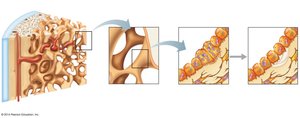
Bone is a dynamic tissue, constantly being deposited and resorbed. Remodeling occurs at periosteal and endosteal surfaces, with osteoblasts depositing and osteoclasts resorbing bone.
Osteoclasts in Bone Resorption
Osteoclasts are large, multinucleated cells that break down bone by secreting hydrochloric acid and lysosomal enzymes. They are derived from hematopoietic stem cells.
Repair of Bone Fractures
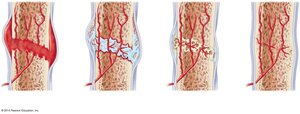
Bone fractures are classified as simple (closed) or compound (open). Healing involves four stages:
Hematoma formation
Fibrocartilaginous callus formation
Bony callus formation
Bone remodeling
Common Types of Fractures
Fracture Type | Description and Comments |
|---|---|
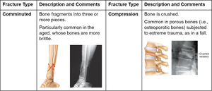
Comminuted | Bone fragments into three or more pieces; common in aged, brittle bones. |
Compression | Bone is crushed; common in porous bones subjected to trauma. |
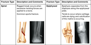
Spiral | Ragged break from excessive twisting forces; common sports fracture. |
Epiphyseal | Epiphysis separates from diaphysis along epiphyseal plate; occurs where cartilage cells are dying. |
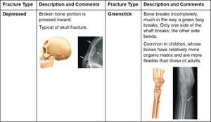
Depressed | Broken bone portion pressed inward; typical of skull fracture. |
Greenstick | Bone breaks incompletely; common in children. |
Disorders of Bones
Osteoporosis
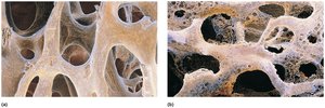
Osteoporosis is characterized by low bone mass, where bone resorption outpaces deposition. It is most common in postmenopausal women.
Osteomalacia and Rickets
Osteomalacia: Inadequate mineralization of bone in adults, leading to soft bones.
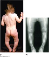
Rickets: Analogous condition in children, resulting in bone deformities.
Osteosarcoma
Osteosarcoma is a malignant bone tumor, most commonly affecting children and young adults.
