 Back
The Skeletal System: Structure, Function, and Classification
Study Guide - Smart Notes
 Tailored notes based on your materials, expanded with key definitions, examples, and context.
The Skeletal System
Overview and Functions
The skeletal system is a complex framework composed of bones, cartilage, joints, and ligaments. It provides the structural support necessary for the human body, facilitates movement, protects internal organs, produces blood cells, and stores essential nutrients such as calcium and fat.
Support: Maintains body shape and supports body weight.
Movement: Acts as levers for muscles to produce movement.
Protection: Shields vital organs (e.g., skull protects the brain, rib cage protects the heart and lungs).
Blood Cell Production: Hematopoiesis occurs in red bone marrow.
Storage: Stores minerals (mainly calcium and phosphate) and lipids (yellow marrow).
Axial and Appendicular Skeleton
Axial Skeleton
The axial skeleton forms the central axis of the body, supporting and protecting the brain, spinal cord, and thoracic organs. It provides attachment points for muscles of the head, neck, and trunk.
Comprised of the skull, vertebral column, and bony thorax (ribs and sternum).
Skull
Cranial bones: Eight bones that enclose the brain and provide muscle attachment.
Facial bones: Fourteen bones that form the face, enclose sense organs, anchor teeth, and provide muscle attachment.
Sutures: Immovable joints connecting skull bones.
Hyoid bone: U-shaped bone in the neck, not directly attached to other bones.
Paranasal Sinuses
Paranasal sinuses are air-filled cavities within certain skull bones that connect to the nasal cavity. They serve to lighten the skull and help warm, cleanse, and filter inhaled air.
Located in the frontal, ethmoid, sphenoid, and maxillary bones.
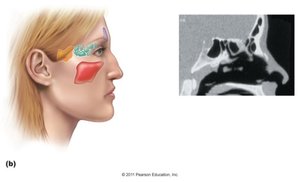
Fetal Skull and Fontanels
Fontanels are fibrous membranes between the bones of the fetal skull. They allow for compression during childbirth and accommodate brain growth.
Anterior, posterior, sphenoidal, and mastoid fontanels.
Vertebral Column
The vertebral column consists of 26 irregular bones that protect the spinal cord, support the trunk, and provide attachment for ribs and muscles.
Cervical vertebrae (7): Support the neck.
Thoracic vertebrae (12): Articulate with ribs.
Lumbar vertebrae (5): Support lower back.
Sacrum: Fusion of five vertebrae, articulates with the ilium.
Coccyx: Tailbone, fusion of four small vertebrae.
Bony Thorax
Sternum: Breastbone in the center of the chest.
Ribs: Twelve pairs; true ribs (1-7), false ribs (8-12), and floating ribs (11-12).
Appendicular Skeleton
The appendicular skeleton includes the bones of the limbs and girdles, enabling movement and manipulation of objects.
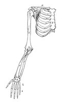
Pectoral girdle: Clavicle and scapula, attaches upper limbs to trunk.
Upper limb: Arm (humerus), forearm (radius and ulna), wrist (carpals), hand (metacarpals and phalanges).
Pelvic girdle: Paired hip bones, attaches lower limbs to the body.
Lower limb: Thigh (femur), knee (patella), leg (tibia and fibula), ankle (tarsals), foot (metatarsals and phalanges).
Cartilage
Types and Functions
Cartilage is an avascular, non-innervated connective tissue composed of chondrocytes in a gel-like matrix. It is surrounded by the perichondrium, which aids in growth and repair.
Hyaline cartilage: Most abundant, strong but flexible; found in articular surfaces, costal cartilages, nose, trachea, and larynx.
Elastic cartilage: Flexible and resilient; found in the external ear and epiglottis.
Fibrocartilage: Strong compression and tension; found in intervertebral discs, pubic symphysis, and menisci of the knee.
Cartilage grows by interstitial (from within) and appositional (from the surface) mechanisms, but has limited repair capacity due to its avascular nature.
Classification of Bones
Bone Shapes
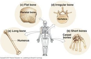
Bones are classified by shape, which relates to their function and location in the body.
Type | Description | Examples |
|---|---|---|
Long bones | Longer than wide | Humerus, femur, phalanges |
Short bones | Cube-shaped, equal length and width | Carpals, tarsals |
Flat bones | Thin, broad | Parietal bone, sternum, ribs |
Irregular bones | Complex shapes | Vertebrae, hip bones |
Macroscopic Anatomy of a Long Bone
Diaphysis: Shaft of the bone, composed of compact bone.
Epiphyses: Expanded ends, composed of spongy bone covered by compact bone and articular cartilage.
Metaphysis: Region between diaphysis and epiphysis, contains the epiphyseal plate (growth plate).
Medullary cavity: Central cavity containing yellow marrow in adults.
Periosteum: Outer fibrous layer and inner cellular layer; site for tendon and ligament attachment, bone growth, and repair.
Endosteum: Lines the medullary cavity and trabeculae of spongy bone; active in bone growth and remodeling.
Microscopic Anatomy of Bone
Bone Cells
Osteoprogenitor cells: Stem cells that differentiate into osteoblasts.
Osteoblasts: Bone-forming cells that secrete bone matrix.
Osteocytes: Mature bone cells maintaining bone tissue, residing in lacunae and communicating via canaliculi.
Osteoclasts: Large, multinucleate cells that resorb bone matrix.
Histology of Compact and Spongy Bone
Osteon: Structural unit of compact bone, consisting of concentric lamellae around a central canal.
Trabeculae: Network of spongy bone filled with marrow.
Bone Growth and Remodeling
Ossification
Intramembranous ossification: Forms flat bones from mesenchymal tissue.
Endochondral ossification: Forms most bones from hyaline cartilage templates.
Bone growth occurs at the epiphyseal plate (interstitial growth) and by appositional growth (increasing diameter). Remodeling is a continuous process involving osteoblasts and osteoclasts, essential for bone strength and mineral homeostasis.
Joints (Articulations)
Classification of Joints
Fibrous joints: Bones joined by dense connective tissue; mostly immovable (e.g., sutures, syndesmoses, gomphoses).
Cartilaginous joints: Bones joined by cartilage; slightly movable (e.g., synchondroses, symphyses).
Synovial joints: Bones separated by a fluid-filled cavity; freely movable (e.g., hinge, ball-and-socket, pivot, saddle, condyloid, plane joints).
Synovial Joint Structure
Articular cartilage: Hyaline cartilage covering bone ends.
Joint cavity: Space containing synovial fluid.
Joint capsule: Fibrous outer layer and synovial membrane.
Reinforcing ligaments: Stabilize the joint.
Bursae and tendon sheaths: Reduce friction.
Types of Synovial Joints
Gliding (plane): Flat surfaces slide past each other (e.g., intercarpal joints).
Hinge: Movement in one plane (e.g., elbow, knee).
Pivot: Rotation (e.g., atlas and axis).
Condyloid: Movement in two planes (e.g., wrist).
Saddle: Angular movement without rotation (e.g., thumb).
Ball-and-socket: All types of movement (e.g., shoulder, hip).
Movements at Synovial Joints
Gliding: Slight movement between flat surfaces.
Angular: Flexion, extension, abduction, adduction, circumduction.
Rotation: Pronation, supination.
Special movements: Inversion, eversion, protraction, retraction, elevation, depression, opposition.
Bone Markings
Bone markings are structural features adapted for specific functions such as muscle attachment, joint formation, or passage of nerves and blood vessels.
Projections: Tuberosity, crest, trochanter, line, tubercle, epicondyle, spine, process, ramus.
Surfaces: Head, facet, condyle, fossa.
Openings: Sinus, meatus, foramen, fissure, notch.
