 Back
The Skeletal System: Structure, Function, and Key Bones
Study Guide - Smart Notes
 Tailored notes based on your materials, expanded with key definitions, examples, and context.
The Skeletal System /
Overview of the Skeletal System
The human skeletal system consists of approximately 206 bones and associated cartilages, organized into two main divisions: the axial and appendicular skeleton. These divisions work together to provide support, protection, and facilitate movement.
Axial Skeleton: Forms the body's longitudinal axis and encases body cavities, protecting vital organs. Includes the skull, vertebral column, and thoracic cage.
Appendicular Skeleton: Comprises the bones of the pectoral and pelvic girdles, as well as the upper and lower limbs. Primarily suited for movement and muscle attachment.
Divisions of the Skeletal System
Skull: 22 bones (8 cranial, 14 facial) encase the brain and form the face.
Vertebral Column: 33 bones; 24 individual vertebrae protect the spinal cord, with the sacrum and coccyx formed by fused vertebrae.
Thoracic Cage: 12 pairs of ribs, sternum, and part of the vertebral column; protects the lungs, heart, and thoracic cavity contents.
Pectoral Girdle: Clavicle and scapula anchor the upper limb to the trunk.
Upper Limb: Arm (humerus), forearm (radius and ulna), wrist and hand (carpals, metacarpals, phalanges).
Pelvic Girdle: Two pelvic bones and sacrum form the pelvis, anchoring the lower limb to the trunk.
Lower Limb: Thigh (femur), leg (tibia and fibula), ankle and foot (tarsals, metatarsals, phalanges).
Bone Markings
Types and Functions of Bone Markings
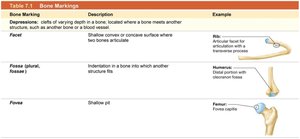
Bones display various surface features known as bone markings, which reflect their structure and function. These markings serve as pathways for blood vessels and nerves, sites for articulations, and attachment points for ligaments and tendons.
Depressions: Clefts of varying depth in a bone, often where a bone meets another structure such as another bone or a blood vessel.
Openings: Enclose delicate structures and allow passage through bones.
Projections: Sites where bones articulate or where ligaments and tendons attach.
Bone Marking | Description | Example |
|---|---|---|
Facet | Shallow concave or convex surface where two bones articulate | Rib: Articular facet for articulation with a transverse process |
Fossa (plural: fossae) | Indentation in a bone into which another structure fits | Humerus: Distal portion with olecranon fossa |
Fovea | Shallow pit | Femur: Fovea capitis |
The Skull
Structure and Organization of the Skull
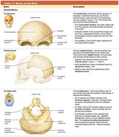
The skull is composed of 22 bones, organized into cranial and facial groups. Cranial bones protect the brain, while facial bones form the framework of the face. Most skull bones are fused at immovable joints called sutures, except for the mandible.
Cranial Bones: 8 bones (frontal, occipital, ethmoid, sphenoid, temporal [paired], parietal [paired])
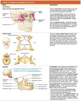
Facial Bones: 14 bones (maxillary, zygomatic, nasal, lacrimal, palatine, inferior nasal conchal [paired], mandible, vomer)
Sinuses: Air-filled, membrane-lined spaces in four bones surrounding the nasal cavity; reduce skull weight and enhance voice resonance.
Key Cranial Bones
Bone | Description |
|---|---|
Frontal Bone | Forms the forehead and superior part of the orbit. |
Parietal Bone | Forms the superior and lateral aspects of the skull. |
Occipital Bone | Forms the posterior and base of the skull. |
Key Facial Bones
Bone | Description |
|---|---|
Maxilla | Forms the upper jaw and part of the orbit. |
Zygomatic Bone | Forms the cheekbones and part of the orbit. |
Mandible | Forms the lower jaw; only movable bone of the skull. |
The Vertebral Column
Structure and Classification
The vertebral column consists of 33 vertebrae, classified by region and function. It protects the spinal cord and supports the head and neck.
Cervical Vertebrae: 7 in the neck
Thoracic Vertebrae: 12 articulate with ribs
Lumbar Vertebrae: 5 in the lower back
Sacral Vertebrae: 5 fused to form the sacrum
Coccygeal Vertebrae: 3–5 fused to form the coccyx
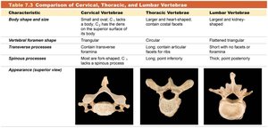
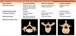
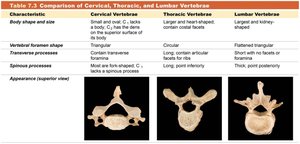
Comparison of Cervical, Thoracic, and Lumbar Vertebrae
Characteristic | Cervical Vertebrae | Thoracic Vertebrae | Lumbar Vertebrae |
|---|---|---|---|
Body shape and size | Small and oval; C1 lacks a body, C2 has the dens on the superior surface of its body | Larger and heart-shaped; contain costal facets | Largest and kidney-shaped |
Vertebral foramen shape | Triangular | Circular | Flattened triangular |
Transverse processes | Most have transverse foramina; C7 lacks transverse foramina | Long; contain articular facets for ribs | Short; no foramina or facets |
Spinous processes | Most are forked; C7 has a spinous process | Long; point inferiorly | Thick; point posteriorly |
Appearance (superior view) |
|
|
|
Study Boost: Memory Aids for Bones
Mnemonic Devices
Pest Of 6: Parietal, Ethmoid, Sphenoid, Temporal, Occipital, Frontal (cranial bones)
Virgil Is Now Making My Pet Zebra Laugh: Vomer, Inferior nasal conchae, Nasal, Mandible, Maxillae, Palatine, Zygomatic, Lacrimal (facial bones)
Breakfast at 7, lunch at 12, dinner at 5: 7 cervical, 12 thoracic, 5 lumbar vertebrae
Clinical Applications
Vertebral Compression Fractures
Vertebral compression fractures reduce the height of a vertebra by more than 20%, often due to trauma or diseases such as osteoporosis. Treatment ranges from conservative management to surgical intervention.
Wedge Fractures: Affect only the anterior portion; common in elderly women.
Burst Fractures: Involve the entire vertebral body; can damage spinal cord and nerves.
Herniated Disc
A tear in the anulus fibrosus allows the nucleus pulposus to protrude, compressing nerve roots and causing pain, numbness, or muscle weakness. Treatments include medication, physical therapy, and surgery.
Summary
The skeletal system is a complex structure essential for protection, support, movement, and mineral storage. Understanding its divisions, bone markings, and clinical relevance is fundamental for students of anatomy and physiology.
