 Back
BackThe Skeletal System: Structure, Function, and Major Components
Study Guide - Smart Notes
 Tailored notes based on your materials, expanded with key definitions, examples, and context.
Overview of the Skeletal System
Structure and Divisions of the Skeletal System
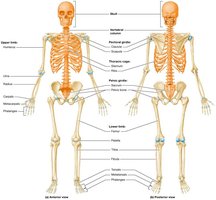
The human skeletal system is composed of approximately 206 bones and associated cartilages, forming the framework that supports and protects the body. The skeleton is divided into two main parts: the axial skeleton and the appendicular skeleton.
Axial Skeleton: Forms the longitudinal axis of the body and includes the skull, vertebral column, and thoracic cage. It primarily functions to protect vital organs such as the brain, spinal cord, heart, and lungs.
Appendicular Skeleton: Comprises the bones of the pectoral girdle, upper limbs, pelvic girdle, and lower limbs. It is mainly involved in movement and muscle attachment.
Bone Markings
Types and Functions of Bone Markings
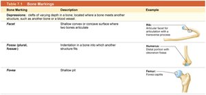
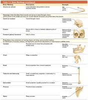
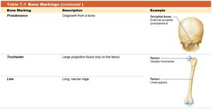
Bones display various surface features known as bone markings, which reflect their functional roles. These markings are classified as depressions, openings, and projections.
Depressions: Serve as pathways for blood vessels and nerves or as sites for articulations between bones.
Openings: Allow the passage of delicate structures through bones.
Projections: Sites where bones articulate or where ligaments and tendons attach.
Bone Marking | Description | Example |
|---|---|---|
Facet | Shallow concave surface where two bones articulate | Rib: Articular facet for articulation with transverse process |
Fossa | Indentation in a bone into which another structure fits | Humerus: Distal portion with olecranon fossa |
Fovea | Shallow pit | Femur: Fovea capitis |
The Skull
Structure and Organization
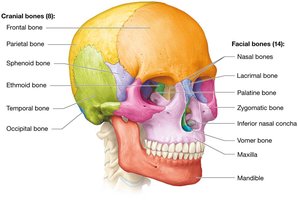
The skull consists of 22 bones organized into cranial and facial groups. Cranial bones (8) protect the brain, while facial bones (14) form the framework of the face. Most skull bones are fused at immovable joints called sutures, except for the mandible.
Cranial Bones: Frontal, parietal, occipital, temporal, ethmoid, and sphenoid bones.
Facial Bones: Maxilla, zygomatic, nasal, lacrimal, palatine, inferior nasal concha, mandible, and vomer.
Cranial Vault and Base
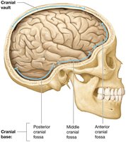
The cranial vault (calvarium) forms the superior portion of the cranial cavity, while the cranial base forms the inferior portion, divided into anterior, middle, and posterior cranial fossae that accommodate the brain.
Cavities of the Skull
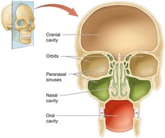
The skull contains several cavities, including the cranial cavity (brain), orbits (eyeballs), nasal cavity (smell), oral cavity (taste), and small cavities for hearing and balance.
Disarticulated Skull
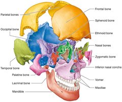
Viewing the skull as a disarticulated model helps in understanding the spatial relationships between individual bones, especially complex ones like the ethmoid and sphenoid.
Orbit and Nasal Cavity
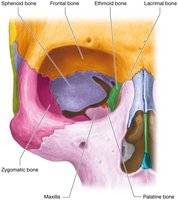
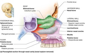
The orbit is formed by seven fused bones and houses the eyeball and associated structures. The nasal cavity is lined with mucous membranes and forms the first part of the respiratory tract.
Fetal Skull and Fontanels
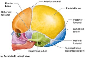
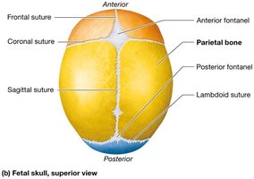
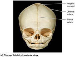
In the fetal skull, bones are not yet fused, and membranous areas called fontanels allow for flexibility during birth and brain growth. Major fontanels include anterior, posterior, sphenoid, and mastoid fontanels.
Hyoid Bone
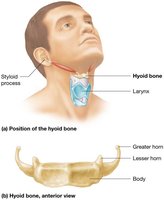
The hyoid bone is a C-shaped bone in the neck that does not articulate with any other bone. It provides attachment points for muscles involved in swallowing and speech.
The Vertebral Column
Structure and Classification
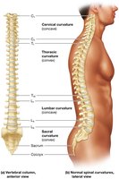
The vertebral column consists of an average of 33 vertebrae, classified by region: 7 cervical, 12 thoracic, 5 lumbar, 5 fused sacral, and 3–5 fused coccygeal vertebrae. The column supports the head, protects the spinal cord, and allows for flexible movement.
Spinal Curvatures
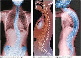
Normal spinal curvatures include primary (thoracic and sacral) and secondary (cervical and lumbar) curves. Abnormal curvatures include scoliosis (lateral), lordosis (exaggerated lumbar), and kyphosis (exaggerated thoracic).
Structure of a Typical Vertebra
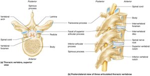
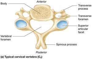
All vertebrae share common features: a body (centrum), vertebral foramen, pedicles, laminae, transverse and spinous processes, and articular processes. These structures provide support, protection, and sites for muscle attachment.
Cervical Vertebrae
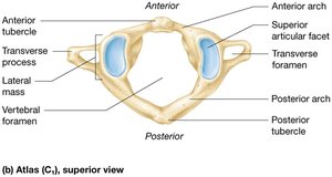
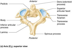
Cervical vertebrae are the smallest and have transverse foramina. The atlas (C1) supports the skull, and the axis (C2) allows for rotation of the head.
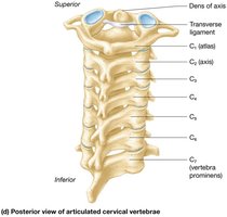
Thoracic and Lumbar Vertebrae
Thoracic vertebrae articulate with ribs and have heart-shaped bodies. Lumbar vertebrae are the largest, supporting the weight of the torso.
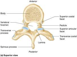
Thoracic Cage
Structure and Function
The thoracic cage consists of the sternum, 12 pairs of ribs, and thoracic vertebrae. It protects the heart and lungs and provides attachment points for muscles involved in respiration.
True Ribs (1–7): Attach directly to the sternum via costal cartilages.
False Ribs (8–12): Do not attach directly to the sternum; ribs 11 and 12 are floating ribs.
Pectoral Girdle and Upper Limb
Pectoral Girdle
The pectoral girdle consists of the clavicle and scapula, supporting the upper limb and providing attachment sites for muscles.
Humerus
The humerus is the only bone of the arm, articulating proximally with the scapula and distally with the radius and ulna.
Forearm: Radius and Ulna
The radius and ulna are the bones of the forearm, connected by an interosseous membrane and articulating with the humerus and carpal bones.
Wrist and Hand
The wrist contains eight carpal bones, while the hand consists of five metacarpals and fourteen phalanges.
Pelvic Girdle and Lower Limb
Pelvis and Pelvic Girdle
The pelvis is formed by the sacrum and two coxal bones (ilium, ischium, and pubis). It supports the weight of the upper body and protects pelvic organs.
Femur and Patella
The femur is the largest bone in the body, articulating with the pelvis at the hip joint and with the tibia at the knee. The patella is a sesamoid bone within the tendon of the quadriceps muscle.
Tibia and Fibula
The tibia is the main weight-bearing bone of the leg, while the fibula provides lateral stability to the ankle.
Ankle and Foot
The ankle contains seven tarsal bones, and the foot contains five metatarsals and fourteen phalanges. The foot's arches help support and distribute body weight during movement.
Study Boost: Mnemonics and Visual Analogies
PEST OF 6: Parietal, Ethmoid, Sphenoid, Temporal, Occipital, Frontal (cranial bones)
Breakfast at 7, lunch at 12, dinner at 5: 7 cervical, 12 thoracic, 5 lumbar vertebrae
Stop Letting The People Touch The Cadaver’s Hand: Scaphoid, Lunate, Triquetrum, Pisiform, Trapezium, Trapezoid, Capitate, Hamate (carpal bones)
Additional info: These mnemonics and analogies are useful for memorizing the names and locations of bones in the skeletal system.
