 Back
BackThe Skeletal System: Structure, Function, and Clinical Correlates
Study Guide - Smart Notes
 Tailored notes based on your materials, expanded with key definitions, examples, and context.
Function of the Skeletal System
Overview of Skeletal Functions
The skeletal system provides the framework for the human body and is essential for movement, protection, and metabolic processes. It consists of bones, cartilage, and ligaments, each contributing to the system's overall function.
Support: Bones form the structural framework that supports the body and cradles soft organs.
Protection: The skeleton protects vital organs (e.g., skull protects the brain, rib cage protects the heart and lungs).
Movement: Muscles attach to bones, and their contraction pulls on bones to produce movement.
Storage: Bones store minerals, primarily calcium and phosphorus.
Parathyroid hormone (PTH): Increases blood calcium levels by stimulating bone resorption.
Calcitonin (CT): Reduces blood calcium levels by promoting bone deposition.
Normal ionized blood calcium: 4.5–5.6 mEq/L.
Hematopoiesis: Blood cell formation occurs in red bone marrow, found in the ends of long bones and flat bones.
Types and Structure of Bones
Classification of Bones
Bones are classified by shape, which relates to their function and location in the body.
Long bones: Longer than they are wide (e.g., humerus, femur).
Short bones: Cube-shaped (e.g., carpals of the wrist).
Flat bones: Thin, flattened, and often curved (e.g., frontal bone of the skull).
Irregular bones: Complex shapes (e.g., vertebrae).
Sesamoid bones: Small, round bones embedded in tendons (e.g., patella).
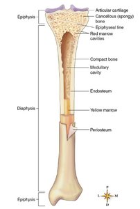
Gross Structure of Long Bones
Long bones have a characteristic structure that supports their function in movement and weight-bearing.
Diaphysis: The shaft; a hollow tube of compact bone providing strong support.
Medullary cavity: Central cavity containing yellow bone marrow (fat storage).
Epiphyses: The ends of the bone; contain red bone marrow for hematopoiesis.
Articular cartilage: Hyaline cartilage covering epiphyses, reducing friction at joints.
Periosteum: Tough membrane covering the bone except at joint surfaces; contains nerves and blood vessels.
Endosteum: Thin membrane lining the medullary cavity.
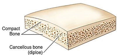
Structure of Flat Bones
Flat bones consist of two layers of compact bone with a spongy bone layer (diploe) in between.
Diploe: The spongy bone layer in flat bones, filled with red or yellow marrow.
Trabeculae: The lattice-like beams of spongy bone.
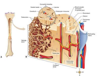
Microscopic Structure of Bone
Compact Bone
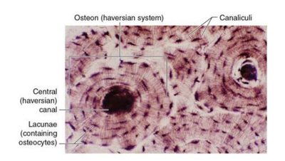
Compact bone is organized into osteons (Haversian systems), which are cylindrical structures that provide strength.
Osteon: The structural unit of compact bone, consisting of concentric lamellae (rings) around a central canal.
Central (Haversian) canal: Contains blood vessels and nerves.
Concentric lamellae: Layers of calcified matrix.
Lacunae: Small spaces containing osteocytes (bone cells).
Canaliculi: Tiny canals connecting lacunae, allowing for nutrient and waste exchange.
Volkmann (transverse) canals: Connect central canals of adjacent osteons.
Bone Formation and Growth
Ossification and Growth
Bone formation (ossification) occurs through two main processes: endochondral ossification (from cartilage) and intramembranous ossification (from connective tissue).
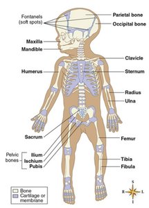
Endochondral ossification: Most bones develop from a cartilage model, especially long bones.
Intramembranous ossification: Flat bones (e.g., skull) develop from fibrous membranes (fontanels in infants).
Epiphyseal plate: Growth in length occurs at the epiphyseal (growth) plate during childhood and adolescence.
Bone Remodeling
Bone is continuously remodeled by the coordinated actions of osteoclasts (bone resorption) and osteoblasts (bone formation). Mechanical stress stimulates bone strengthening at sites of muscle attachment.
Bone Disorders: Osteoporosis
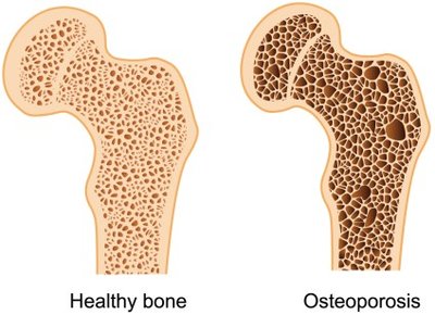
Osteoporosis
Osteoporosis is an age-related disease characterized by decreased bone mass and increased fracture risk due to excessive osteoclast activity and loss of calcified matrix.
Risk factors: Advanced age, female gender, sedentary lifestyle, low calcium/vitamin D intake, genetics, certain medications, and hormonal imbalances.
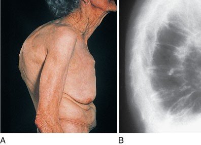
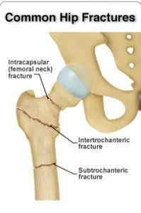
Clinical features: Pathologic fractures, spinal deformities (Dowager’s hump), loss of height, and pain.
Diagnosis: Bone density testing (DXA scan), X-ray imaging.
Management: Weight-bearing exercise, fall prevention, calcium/vitamin D supplementation, lifestyle modifications.
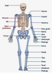
Divisions of the Skeleton
Axial and Appendicular Skeleton
The human skeleton is divided into the axial and appendicular skeletons.
Axial skeleton: Skull, vertebral column, thoracic cage (ribs and sternum), and hyoid bone.
Appendicular skeleton: Upper and lower limbs, pectoral (shoulder) girdle, and pelvic (hip) girdle.
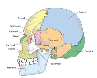
Skull and Associated Structures
Skull Bones
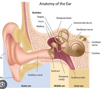
The skull consists of cranial and facial bones, as well as the middle ear bones.
Cranium: 8 fused bones protecting the brain.
Facial bones: 14 bones forming the face.
Middle ear bones (ossicles): Malleus, incus, and stapes transmit sound vibrations.
Vomer: Forms the posterior part of the nasal septum.
Zygomatic bone: Cheekbone.
Mastoid process: Part of the temporal bone.
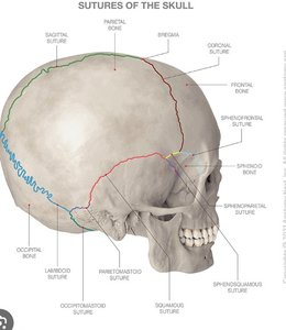
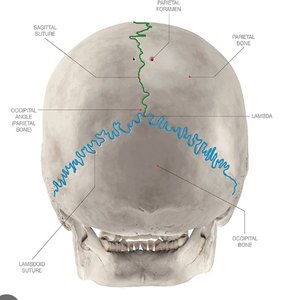
Sutures of the Skull
Sutures are immovable joints connecting the bones of the skull.
Lambdoidal suture: Parietal bones to occipital bone.
Squamous suture: Parietal to temporal and sphenoid bones.
Coronal suture: Parietal to frontal bone.
Sagittal suture: Between parietal bones.
Middle Ear Bones
The ossicles (malleus, incus, stapes) are the smallest bones in the body and transmit sound from the tympanic membrane to the inner ear.
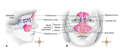
Paranasal Sinuses
Sinuses are air-filled cavities in the skull that lighten its weight and enhance voice resonance. The four pairs are frontal, maxillary, sphenoid, and ethmoid sinuses.
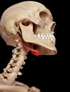
Hyoid Bone
The hyoid bone is a U-shaped bone in the neck that does not articulate with any other bone. It supports the tongue and is associated with swallowing.
Vertebral Column
Regions and Structure
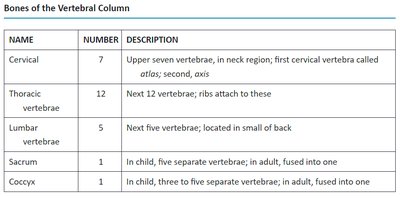
The vertebral column consists of 33 vertebrae in childhood, which fuse to 26 in adulthood. It is divided into cervical, thoracic, lumbar, sacral, and coccygeal regions.
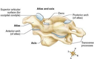
Cervical (7): Neck region; includes atlas (C1) and axis (C2).
Thoracic (12): Chest region; ribs attach here.
Lumbar (5): Lower back.
Sacrum (1): Five fused vertebrae.
Coccyx (1): Three to five fused vertebrae (tailbone).
NAME | NUMBER | DESCRIPTION |
|---|---|---|
Cervical | 7 | Upper seven vertebrae; first cervical vertebra called atlas; second, axis |
Thoracic | 12 | Next 12 vertebrae; ribs attach to these |
Lumbar | 5 | Next five vertebrae; located in small of back |
Sacrum | 1 | In child, five separate vertebrae; in adult, fused into one |
Coccyx | 1 | In child, three to five separate vertebrae; in adult, fused into one |
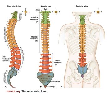
Abnormal Spinal Curvatures
Scoliosis: Lateral curvature of the spine.
Kyphosis: Exaggerated thoracic curvature.
Lordosis: Exaggerated lumbar curvature.
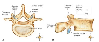
Vertebral Anatomy
Spinous process: Posterior projection for muscle attachment.
Transverse process: Lateral projections for muscle and ligament attachment.
Vertebral foramen: Canal for the spinal cord.
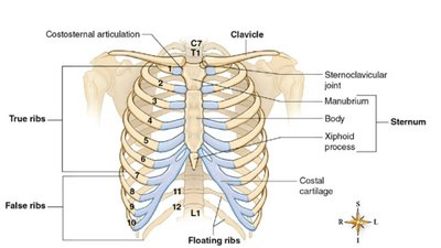
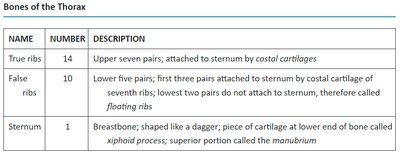
Thorax
Structure of the Thorax
The thorax consists of the sternum, ribs, and thoracic vertebrae, forming the rib cage that protects the heart and lungs.
NAME | NUMBER | DESCRIPTION |
|---|---|---|
True ribs | 14 | Upper seven pairs; attached to sternum by costal cartilages |
False ribs | 10 | Lower five pairs; first three pairs attached to sternum by costal cartilage of seventh ribs; lowest two pairs do not attach to sternum, called floating ribs |
Sternum | 1 | Breastbone; shaped like a dagger; piece of cartilage at lower end called xiphoid process; superior portion called manubrium |
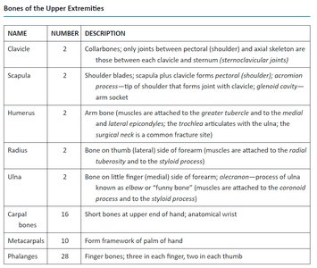
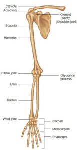
Appendicular Skeleton
Upper Extremity and Pectoral Girdle
Clavicle: Collarbone; connects arm to trunk.
Scapula: Shoulder blade.
Humerus: Upper arm bone.
Radius and Ulna: Forearm bones.
Carpals, Metacarpals, Phalanges: Bones of the wrist, hand, and fingers.
NAME | NUMBER | DESCRIPTION |
|---|---|---|
Clavicle | 2 | Collarbones; only joints between pectoral (shoulder) and axial skeleton are those between each clavicle and sternum (sternoclavicular joints) |
Scapula | 2 | Shoulder blades; scapula plus clavicle forms pectoral (shoulder) girdle; glenoid cavity—arm socket |
Humerus | 2 | Arm bone |
Radius | 2 | Bone on thumb (lateral) side of forearm |
Ulna | 2 | Bone on little finger (medial) side of forearm |
Carpal bones | 16 | Short bones at upper end of hand, anatomical wrist |
Metacarpals | 10 | Form framework of palm of hand |
Phalanges | 28 | Finger bones; three in each finger, two in each thumb |
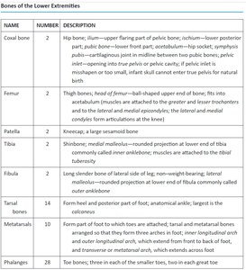
Lower Extremity and Pelvic Girdle
Coxal bones: Hip bones; form pelvic girdle.
Femur: Thigh bone; longest and strongest bone in the body.
Patella: Kneecap; a sesamoid bone.
Tibia and Fibula: Bones of the lower leg.
Tarsals, Metatarsals, Phalanges: Bones of the ankle, foot, and toes.
NAME | NUMBER | DESCRIPTION |
|---|---|---|
Coxal bone | 2 | Hip bone; ilium, ischium, pubis |
Femur | 2 | Thigh bone |
Patella | 2 | Kneecap |
Tibia | 2 | Shinbone |
Fibula | 2 | Lower side of leg |
Tarsal bones | 14 | Short bones of ankle |
Metatarsals | 10 | Form framework of foot |
Phalanges | 28 | Toe bones |
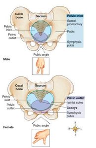
Sexual Dimorphism in the Skeleton
Male and female skeletons differ, especially in the pelvis, to accommodate childbirth in females.
Male skeleton: Generally larger, pelvis is narrow, pelvic inlet is smaller, pubic angle is more acute.
Female skeleton: Broader pelvis, wider pelvic inlet, larger pubic angle.
Cartilage
Structure and Function
Chondrocytes: Cartilage cells embedded in a gel-like matrix.
Matrix: Firm, flexible, and avascular (lacks blood vessels).
Repair: Cartilage repairs slowly due to lack of blood supply.
Joints (Articulations)
Classification of Joints
Synarthroses: Immovable joints (e.g., cranial sutures).
Amphiarthroses: Slightly movable joints (e.g., symphysis pubis, intervertebral discs).
Diarthroses (synovial joints): Freely movable joints (e.g., hip, knee, shoulder).
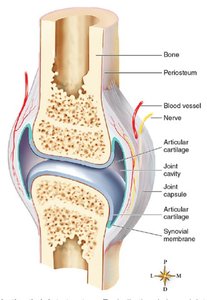
Structure of Synovial Joints
Joint capsule: Encloses the joint cavity.
Synovial membrane: Secretes synovial fluid for lubrication.
Articular cartilage: Covers bone surfaces at joints.
Ligaments: Reinforce and stabilize joints.
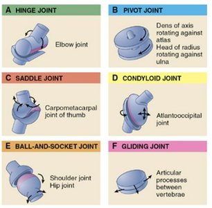
Types of Synovial Joints and Movements
Ball-and-socket: Shoulder and hip; movement in all planes.
Hinge: Elbow and knee; flexion and extension.
Pivot: Rotation (e.g., atlas and axis).
Saddle: Thumb joint.
Gliding: Intercarpal joints.
Condyloid: Wrist joint.
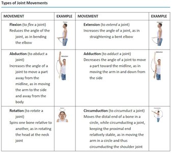
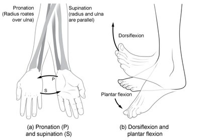
Types of Joint Movements
MOVEMENT | EXAMPLE | MOVEMENT | EXAMPLE |
|---|---|---|---|
Flexion | Bending the elbow | Extension | Straightening the elbow |
Abduction | Raising arm away from body | Adduction | Lowering arm toward body |
Rotation | Turning head side to side | Circumduction | Moving arm in a circle |
Clinical Correlate: Osteoarthritis
Osteoarthritis
Osteoarthritis is a degenerative joint disease affecting weight-bearing joints and hands, often due to wear and tear. It is more common in older adults and those with obesity or joint overuse.
Symptoms: Stiffness, crepitus, decreased range of motion, pain (worse with movement), joint swelling, and deformities.
Treatment: Pain control (acetaminophen, NSAIDs), physical therapy, weight loss, intra-articular steroid injections, and joint replacement if necessary.
Additional info: This guide covers the essential structure, function, and clinical aspects of the skeletal system, suitable for ANP college-level study.
