 Back
BackThe Skeletal System: Structure, Function, and Bone Physiology
Study Guide - Smart Notes
 Tailored notes based on your materials, expanded with key definitions, examples, and context.
The Skeletal System
Components of the Skeletal System
The skeletal system is a complex organ system composed of bones, joints, cartilage, ligaments, and tendons. Each component plays a distinct role in providing structure, movement, and protection for the body.
Bones: Rigid organs that form the framework of the body.
Joints: Sites where two bones articulate, allowing movement.
Cartilage: Flexible connective tissue found between bones and joints.
Ligaments: Connect bone to bone, stabilizing joints.
Tendons: Connect muscle to bone, enabling movement.
Cartilage Types and Functions
Cartilage is essential for flexibility and cushioning in the skeletal system. It is avascular and surrounded by the perichondrium, which limits outward expansion.
Hyaline cartilage: Provides support with flexibility; found in articular, costal, respiratory, and nasal regions.
Elastic cartilage: Contains more elastic fibers; allows stretching; found in external ears and epiglottis.
Fibrocartilage: Provides cushioning and strength; found in intervertebral discs, meniscus, and pubic symphysis.
Bone Structure and Classification
Bones as Organs
Bones are considered organs because they contain multiple tissue types and perform various functions, including structure, movement, protection, blood cell production, mineral storage, and muscle anchoring.
Axial vs. Appendicular Skeleton
The human skeleton consists of 206 bones, divided into two main groups:
Axial skeleton: Forms the longitudinal axis (skull, vertebral column, thoracic cage).
Appendicular skeleton: Includes limbs and girdles attaching them to the axial skeleton.
Classification of Bones by Shape
Bones are classified based on their shape and internal structure:
Long bones: Longer than wide, with a shaft and enlarged ends (e.g., humerus, femur).
Short bones: Cube-shaped, mostly spongy bone; includes sesamoid bones (e.g., talus).
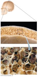
Flat bones: Thin, often curved, with a spongy layer sandwiched between two compact layers (e.g., skull, ribs).
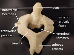
Irregular bones: Complex shapes that do not fit other categories (e.g., vertebrae).
Gross Anatomy of Long Bones
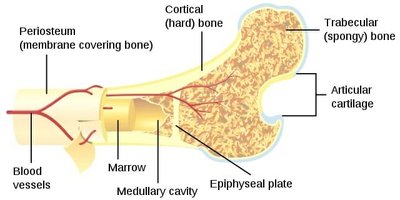
Long bones have distinct anatomical regions:
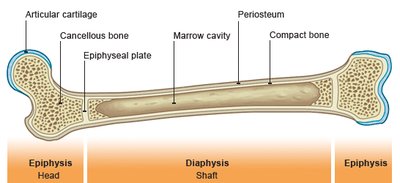
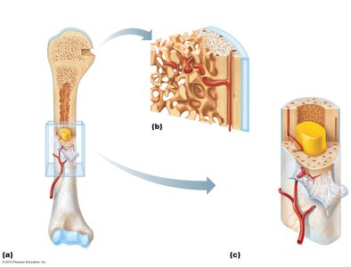
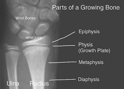
Diaphysis: Shaft, composed of compact bone covered by periosteum.
Epiphysis: Ends, filled with spongy bone and covered with articular cartilage.
Epiphyseal plate: Hyaline cartilage responsible for lengthwise growth; becomes epiphyseal line in adults.
Medullary cavity: Central cavity for marrow storage (red in children, yellow in adults).
Endosteum: Membrane lining the inner surface of the bone.
Bone Markings
Bones display various markings that serve as landmarks for muscle attachment, joint formation, and passage of nerves and blood vessels.
Projections (bulges): Heads, trochanters, spines.
Depressions and openings: Fossa, sinuses, foramina, grooves.
Structure of Short Bones
Short bones consist of a sandwich of spongy bone between compact layers, lacking a shaft or epiphyses. Bone marrow is found between trabeculae, but there is no marrow cavity.
Microscopic Anatomy of Bone
Osteon (Haversian System)
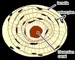
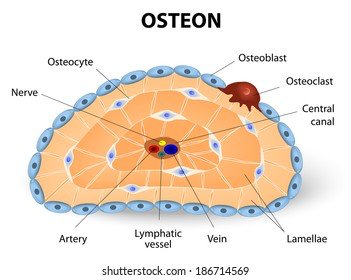
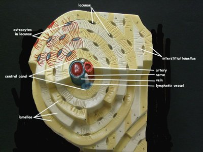
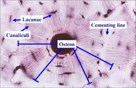
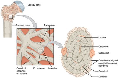
The osteon is the structural and functional unit of compact bone. Osteocytes reside in lacunae, which are arranged in concentric lamellae around a central (Haversian) canal.
Lamellae: Concentric rings of bone matrix.
Central canal: Contains blood vessels and nerves.
Canaliculi: Tiny channels connecting lacunae, facilitating nutrient and waste exchange.
Bone Vascularization
Central canals run lengthwise, carrying blood vessels and nerves. Perforating (Volkmann's) canals run at right angles, connecting the exterior to the interior of bone.
Bone Formation and Growth
Ossification Processes
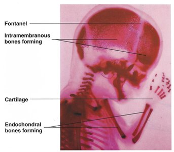
Bone formation (ossification) begins at 8 weeks gestation and occurs via two mechanisms:
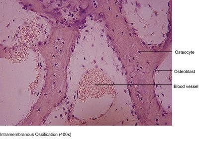
Intramembranous ossification: Forms cranial bones and clavicles from fibrous membranes.
Endochondral ossification: Forms most bones from hyaline cartilage templates.
Epiphyseal Plate and Bone Growth
The epiphyseal plate is the site of active ossification, where chondrocytes divide and push the epiphyses apart, lengthening the bone. This plate calcifies in early adulthood.
Factors Affecting Bone Growth
Bone growth is influenced by genetics, nutrition, and hormones:
Genetics: Most influential factor.
Nutrition: Calcium, Vitamin D (for Ca2+ absorption), and Vitamin C (for collagen synthesis).
Hormones: Growth hormone, thyroid hormone, and sex hormones regulate growth and epiphyseal closure.
Bone Matrix Composition
Organic and Inorganic Components
The bone matrix consists of both organic and inorganic materials:
Organic: Cells, collagen, ground substance (1/3 dry weight).
Inorganic: Hydroxyapatite (calcium phosphate salt, 85%), calcium carbonate (10%), other minerals (2/3 dry weight).
Collagen provides flexibility and strength, while minerals confer rigidity.
Bone Remodeling and Homeostasis
Bone Remodeling
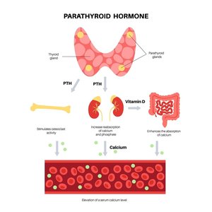
Bones are continuously remodeled in response to calcium levels and mechanical forces. Parathyroid hormone (PTH) regulates calcium homeostasis:
If blood calcium is low, PTH stimulates osteoclasts to break down bone and release Ca2+.
If blood calcium is high, osteoblasts deposit Ca2+ onto bone.
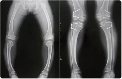
Rickets is a disorder where bones fail to calcify, usually due to lack of calcium or Vitamin D.
Bone Fractures and Healing
Types of Bone Fractures
Bone fractures are classified by their characteristics:
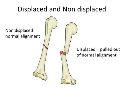
Displaced vs. Non-displaced: Displaced fractures are misaligned; non-displaced remain in normal alignment.
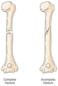
Complete vs. Incomplete: Complete fractures penetrate the entire bone; incomplete affect only part of the bone.
Linear vs. Transverse: Linear fractures run parallel to the bone's axis; transverse are perpendicular.
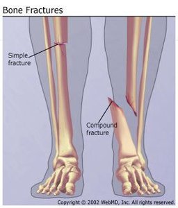
Simple (closed) vs. Compound (open): Simple fractures do not penetrate the skin; compound fractures do.
Bone Healing Process
Bone healing occurs in four stages:
Hematoma formation (blood-filled swelling).
Fibrocartilage callus formation from endosteum and periosteum cells.
Bony callus formation, replacing fibrocartilage with spongy bone.
Bone remodeling to restore original shape and structure.
Bones and Aging
Bone Mass and Osteoporosis
Bone mass peaks around age 30 and declines with age, especially in women after menopause due to decreased estrogen. Osteoporosis results from an imbalance between bone resorption and formation, leading to fragile bones.
Causes: Inadequate calcium, overproduction of PTH, overactive osteoclasts.
Treatments: Nutritional supplements, weight-bearing exercise, hormone replacement therapy.
Blood Calcium Homeostasis
Calcium Regulation
Calcium ions are vital for nerve signaling, muscle contraction, blood clotting, and cell division. Blood calcium is tightly regulated, primarily by PTH.
Increase by 30% disrupts nerve and muscle activity.
Decrease by 35% causes overexcited neurons and convulsions.
Practice Question: Osteoporosis Therapy
Osteoporosis is caused by an imbalance between bone resorption and formation. A therapeutic drug that inhibits osteoclast activity and enhances osteoblast activity would be useful for treatment.
Correct answer: d. A therapeutic drug that inhibits osteoclast activity would be useful. One that enhances the bone formation activity of osteoblasts may also be a good strategy.
Summary Table: Common Types of Bone Fractures
Fracture Type | Description | Comment |
|---|---|---|
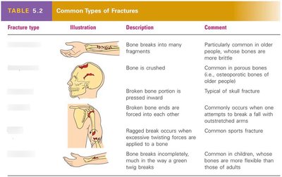
Comminuted | Bone breaks into many fragments | Common in older people, whose bones are more brittle |
Compression | Bone is crushed | Common in porous bones (e.g., osteoporotic bones of older people) |
Depressed | Broken bone portion is pressed inward | Typical of skull fracture |
Impacted | Broken bone ends are forced into each other | Occurs when one attempts to break a fall with outstretched arms |
Spiral | Ragged break occurs when excessive twisting forces are applied | Common sports fracture |
Greenstick | Bone breaks incompletely, much like a green twig breaks | Common in children, whose bones are more flexible than those of adults |
