 Back
BackThe Skeletal System: Structure, Function, and Disorders
Study Guide - Smart Notes
 Tailored notes based on your materials, expanded with key definitions, examples, and context.
The Skeletal System
Introduction to the Skeletal System
The skeletal system is a complex organ system that provides the framework for the human body. It consists of approximately 206 bones in adults, as well as cartilages, ligaments, joints, and other connective tissues that stabilize or connect bones. In infants, the number of bones is higher due to incomplete fusion.
Protection: Shields vital organs such as the brain, heart, and lungs.
Support: Provides structural support for the entire body.
Blood Cell Production: Hematopoiesis occurs in red bone marrow.
Storage: Stores lipids (in yellow marrow) and minerals (mainly calcium and phosphate).
Leverage: Acts as levers for muscles to produce movement.
Organization of the Skeleton
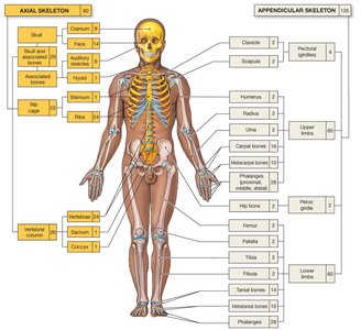
The skeleton is divided into two main parts: the axial skeleton and the appendicular skeleton.
Axial Skeleton: Includes the skull, thorax, and vertebral column; forms the longitudinal axis of the body.
Appendicular Skeleton: Includes the bones of the limbs and the pectoral and pelvic girdles, which attach the limbs to the axial skeleton.
Bone Classifications
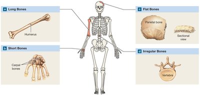
Bones are classified by shape, which is closely related to their function.
Flat Bones: Thin and broad (e.g., parietal bone).
Long Bones: Longer than they are wide (e.g., humerus).
Short Bones: About as long as they are wide (e.g., carpal bones).
Irregular Bones: Complex shapes (e.g., vertebrae).
Sesamoid Bones: Small, round, and embedded in tendons (e.g., patella).
Bone markings include elevations or projections for tendon and ligament attachment, and depressions, grooves, or tunnels for blood vessels or nerve passages.
Bone Structure and Tissue
Structure of a Long Bone
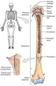
Long bones have a characteristic structure that supports their function in movement and weight-bearing.
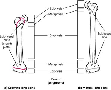
Diaphysis: The shaft of the bone.
Epiphysis: The ends of the bone, often wider than the shaft.
Metaphysis: The region between the diaphysis and epiphysis.
Marrow Cavity: Contains bone marrow (red or yellow).
Periosteum: A dense layer of vascular connective tissue enveloping the bone except at the surfaces of the joints.
Types of Bone Tissue
Bone tissue is specialized connective tissue composed of cells and an extracellular matrix. There are two main types:
Compact Bone: Dense and forms the outer layer of bones; composed of osteons; resists stress in a specific direction.
Spongy Bone: Found inside bones; consists of trabeculae; reduces bone weight and provides strength; spaces filled with marrow.
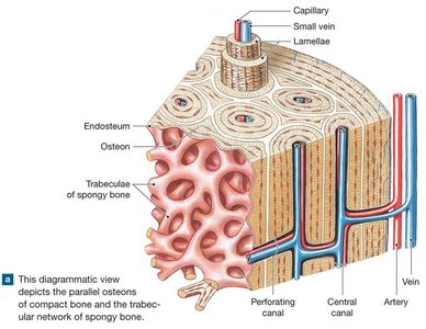
Microscopic Structure: The Osteon
The osteon (Haversian system) is the fundamental functional unit of compact bone.
Central Canal: Contains blood vessels and nerves.
Lamellae: Concentric rings of bone matrix.
Lacunae: Small spaces containing osteocytes.
Canaliculi: Tiny canals connecting lacunae for nutrient and waste exchange.
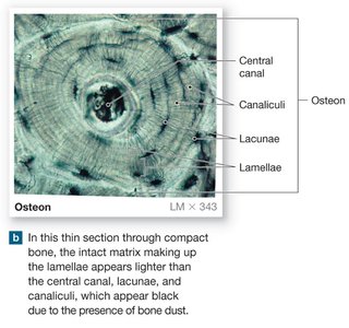
Bone Cells and Development
Bone and Cartilage Cells
Bone and cartilage tissues contain specialized cells:
Chondrocytes: Mature cartilage cells in lacunae.
Chondroblasts: Young, active cartilage cells that produce matrix.
Osteocytes: Mature bone cells that maintain bone matrix.
Osteoblasts: Bone-forming cells; synthesize new bone matrix.
Osteoclasts: Bone-resorbing cells; break down bone matrix.
Bone Development (Ossification)
Bone formation begins in the embryo and continues into adulthood. There are two main processes:
Intramembranous Ossification: Bone develops directly from mesenchymal tissue (e.g., flat bones of the skull).
Endochondral Ossification: Bone replaces a cartilage model (e.g., long bones).
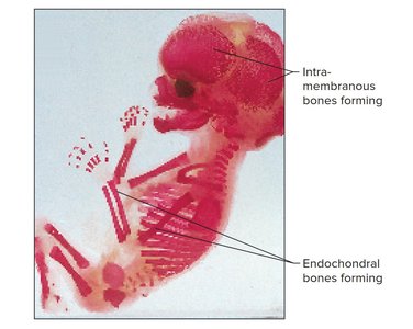
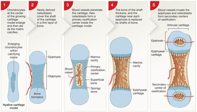
Bone Growth
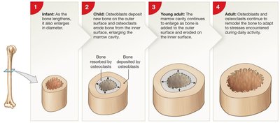
Bones grow in length and diameter through two processes:
Interstitial Growth: Growth in length at the epiphyseal plate; closure results in the epiphyseal line.
Appositional Growth: Growth in diameter/thickness by adding new bone to the surface.
Bone Remodeling and Homeostasis
Bone Remodeling
Bone is a dynamic tissue that undergoes continuous remodeling. About 3-5% of bone calcium is exchanged each year. Remodeling involves:
Osteocytes: Maintain bone matrix.
Osteoclasts: Remove bone matrix. Matriz osea
Osteoblasts: Build new bone matrix.
Factors affecting remodeling include nutrition (vitamins A, C, D), exercise, and hormones.
Hormonal Regulation of Bone
Several hormones regulate bone growth and calcium homeostasis:
Growth Hormone (GH): Stimulates growth at the epiphyseal plate.
Thyroid Hormones (T3, T4): Regulate metabolism.
Sex Hormones (Estrogen, Testosterone): Stimulate osteoblasts and influence epiphyseal plate closure.
Calcitonin: Promotes calcium deposition in bone, lowering blood calcium levels.
Parathyroid Hormone (PTH): Increases blood calcium by stimulating osteoclast activity.
Skeletal Disorders
Atypical Skeletal Growth
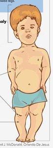
Achondroplasia: Slow growth of epiphyseal cartilage; results in short limbs but normal trunk size.
Gigantism: Overproduction of growth hormone before puberty; results in excessive height.
Acromegaly: Overproduction of growth hormone after epiphyseal closure; bones thicken, especially in the face, jaw, and hands.

Osteopenia and Osteoporosis
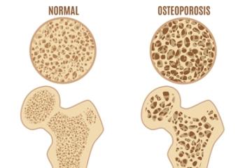
Osteopenia: Age-related loss of bone mass; often involves loss of cartilage from joints.
Osteoporosis: Severe loss of bone density and mass, increasing fracture risk.
Bone Fractures
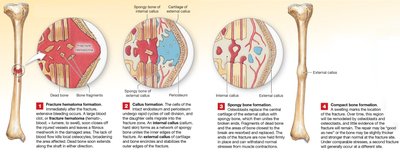
A fracture is a crack or break in a bone. Fractures are classified as closed (simple) or open (compound).
Joints (Articulations)
Classification of Joints
Joints are classified by their degree of movement and structure:
Synarthrotic: No movement (e.g., sutures of the skull).
Amphiarthrotic: Slight movement (e.g., symphysis of the pubic bone).
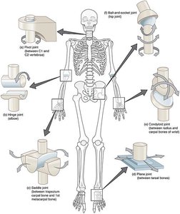
Diarthrotic (Synovial): Free movement (e.g., knee, elbow).
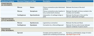
Functional Category | Structural Category and Type | Description | Example |
|---|---|---|---|
Synarthrosis (No Movement) | Fibrous: Suture, Gomphosis; Cartilaginous: Synchondrosis | Fibrous connections or cartilage bridge | Skull sutures, teeth sockets, epiphyseal cartilages |
Amphiarthrosis (Little Movement) | Fibrous: Syndesmosis; Cartilaginous: Symphysis | Ligament or fibrocartilage pad | Between tibia and fibula, pubic symphysis |
Diarthrosis (Free Movement) | Synovial | Joint capsule with synovial fluid | Knee, elbow, shoulder |
Synovial Joints
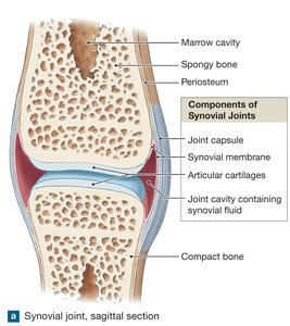
Synovial joints are the most movable type of joint in the body. They have a joint capsule, synovial membrane, articular cartilage, and a joint cavity filled with synovial fluid.
Synovial Fluid: Lubricates the joint, reduces friction, and nourishes cartilage.
Articular Cartilage: Covers bone surfaces, absorbs shock, and reduces friction.
Disorders of Joints: Arthritis
Arthritis refers to over 100 different types of inflammatory or degenerative diseases that damage joints. The most common types include:
Rheumatoid Arthritis: Chronic autoimmune disorder; the body attacks the synovial membrane.
Osteoarthritis: Degenerative joint disease due to wear and tear.
