 Back
BackThe Skeletal System: Structure, Function, and Anatomy
Study Guide - Smart Notes
 Tailored notes based on your materials, expanded with key definitions, examples, and context.
The Skeletal System
Introduction to Bone and Skeletal Tissue
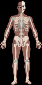
The skeletal system is composed of bones and associated connective tissues. Bones are dynamic, living organs that respond to their environment and perform several essential functions in the human body.
Support: Bones provide a rigid framework that supports the body and maintains its shape.
Protection: Bones protect soft internal organs, such as the brain, heart, and lungs.
Movement: Bones act as levers for muscles, enabling movement.
Mineral Storage: Bones store minerals, primarily calcium and phosphate, which can be released into the bloodstream as needed.
Blood Cell Production: Bones house bone marrow, which produces blood cells (hematopoiesis).
Fat Storage: Yellow bone marrow stores triglycerides (fat).



Types of Bones
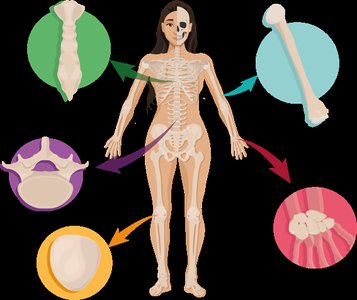
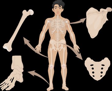
Bones are classified based on their shapes and structures, each adapted for specific functions.
Long Bones: Longer than they are wide, with a shaft and two ends (e.g., femur, humerus).
Short Bones: Cube-shaped, nearly equal in length and width (e.g., carpals, tarsals).
Flat Bones: Thin, flattened, and usually curved (e.g., sternum, ribs, cranial bones).
Irregular Bones: Complex shapes that do not fit other categories (e.g., vertebrae, pelvis).
Sesamoid Bones: Develop within tendons (e.g., patella); number varies among individuals.
Gross Anatomy of Bone: Compact and Spongy Bone
Bone tissue is organized into two main structural types: compact bone and spongy bone.
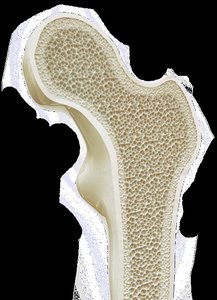
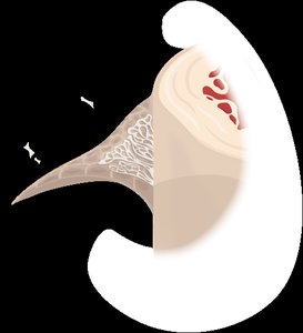
Compact Bone: Dense, solid, and forms the outer layer of all bones; provides strength and protection.
Spongy Bone (Cancellous Bone): Porous, lattice-like structure found inside bones; reduces weight and contains marrow.
Analogy: Compact bone is like a brick wall (strong and solid), while spongy bone is like scaffolding (lightweight but supportive).

Gross Anatomy of Bone: Periosteum and Endosteum
Bones are covered and lined by specialized connective tissues:
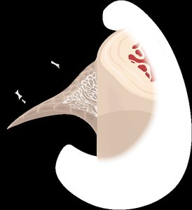
Periosteum: A double-layered membrane covering the external surface of bones, containing blood vessels, nerves, and bone stem cells. The outer fibrous layer is dense irregular connective tissue; the inner osteogenic layer contains osteoprogenitor cells.
Endosteum: A thin membrane lining the internal surfaces of bones, including the medullary cavity and trabeculae of spongy bone; similar in composition to the osteogenic layer of the periosteum.
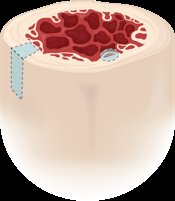
Gross Anatomy of Bone: Bone Marrow
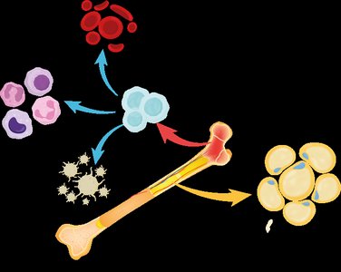
Bone marrow is a soft tissue found in the cavities of bones and exists in two forms:
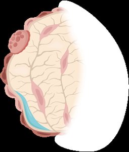
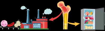
Red Marrow: Site of hematopoiesis (production of blood cells); found mainly in spongy bone of adults and in most bones of infants.
Yellow Marrow: Stores fat (triglycerides); found in the medullary cavity of long bones in adults. Can revert to red marrow if necessary.

Gross Anatomy of Bones: Structure of a Long Bone
Long bones have a characteristic structure:
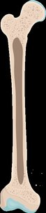
Epiphysis: The expanded ends of the bone, covered with articular cartilage.
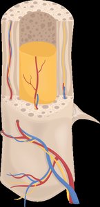
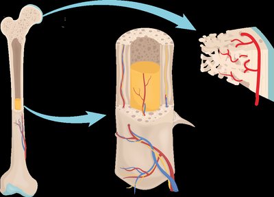
Diaphysis: The shaft of the bone, containing the medullary cavity filled with yellow marrow.
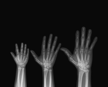
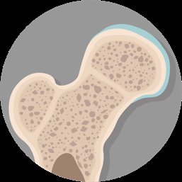
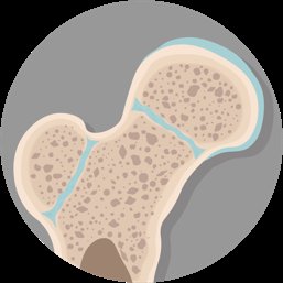
Metaphysis: The region where the diaphysis and epiphysis meet; contains the epiphyseal plate (growth plate) in children and adolescents, which becomes the epiphyseal line in adults.
Gross Anatomy of Bones: Nerves and Blood Supply
Bones are highly vascularized and innervated:
Nutrient Foramen: Small openings in the diaphysis for blood vessels and nerves to enter and exit the bone.
Nutrient Artery and Vein: Supply and drain blood from the medullary cavity.
Smaller Foramina: Located in the metaphysis and epiphysis for additional blood supply.
Microscopic Anatomy of Bones: Bone Matrix
The extracellular matrix of bone consists of two main components:
Inorganic Matrix: Hydroxyapatite crystals (calcium and phosphate) provide hardness and strength; about 65% of bone mass.
Organic Matrix (Osteoid): Collagen fibers and ground substance provide flexibility and tensile strength; about 35% of bone mass.
Equation for Hydroxyapatite: $\mathrm{Ca_{10}(PO_4)_6(OH)_2}$
Microscopic Anatomy of Bones: Bone Cells
Bone tissue contains several types of cells, each with specialized functions:
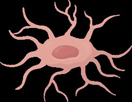
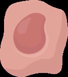
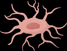
Osteoprogenitor (Osteogenic) Cells: Stem cells that differentiate into osteoblasts.
Osteoblasts: Bone-forming cells that secrete osteoid (organic matrix).
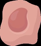
Osteocytes: Mature bone cells that maintain the bone matrix; reside in lacunae and communicate via canaliculi.
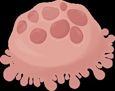
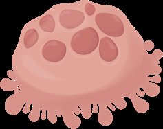
Osteoclasts: Large, multinucleated cells that break down bone matrix (osteolysis) for remodeling and calcium release.
Microscopic Anatomy of Bones: The Osteon
The osteon (Haversian system) is the structural unit of compact bone:
Central Canal: Contains blood vessels and nerves; runs parallel to the long axis of the bone.
Lamellae: Concentric rings of bone matrix around the central canal.
Lacunae: Small spaces containing osteocytes.
Canaliculi: Tiny channels connecting lacunae for nutrient and signal exchange.
Perforating (Volkmann's) Canals: Run perpendicular to central canals, connecting blood and nerve supply.
Microscopic Anatomy of Bones: Trabeculae
Spongy bone is composed of trabeculae, which are small, needle-like pieces of bone aligned along lines of stress. Trabeculae contain lamellae, osteocytes, and canaliculi but lack central canals.
Introduction to the Skeleton
The human skeleton consists of 206 named bones, divided into two main divisions:
Axial Skeleton: Skull, vertebral column, and rib cage (80 bones); provides support and protection for internal organs.
Appendicular Skeleton: Limbs and girdles (126 bones); enables movement.
The Skull
The skull is a complex structure composed of cranial and facial bones, associated bones, and cavities/sinuses. It protects the brain, forms facial structure, provides openings for air and food, anchors teeth, and houses sensory organs.
The Spine (Vertebral Column)
The vertebral column consists of 24 vertebrae, the sacrum, and the coccyx. It is divided into cervical, thoracic, lumbar, sacral, and coccygeal regions, each with characteristic vertebrae and curvatures. Intervertebral discs provide cushioning and flexibility.
The Thoracic Cage
The thoracic cage includes the ribs, sternum, and thoracic vertebrae. It protects the thoracic organs and supports respiration. Ribs are classified as true, false, or floating based on their attachment to the sternum.
The Pectoral Girdle and Upper Limb
The pectoral girdle (clavicle and scapula) attaches the upper limbs to the axial skeleton. The upper limb consists of the humerus, radius, ulna, carpals, metacarpals, and phalanges.
The Pelvic Girdle and Lower Limb
The pelvic girdle (coxal bones) attaches the lower limbs to the axial skeleton. The lower limb includes the femur, patella, tibia, fibula, tarsals, metatarsals, and phalanges. The structure of the pelvis differs between males and females, with adaptations for childbirth in females.
Introduction to Joints
Joints (articulations) are points of contact between bones or between bone and teeth. They are classified by structure (fibrous, cartilaginous, synovial) and function (synarthrosis, amphiarthrosis, diarthrosis). Synovial joints are the most movable and have unique features such as a synovial cavity, articular cartilage, and an articular capsule.
Classification of Joints
Fibrous Joints: Bones joined by dense connective tissue; little or no movement (e.g., sutures, syndesmoses, gomphoses).
Cartilaginous Joints: Bones joined by cartilage; limited movement (e.g., synchondroses, symphyses).
Synovial Joints: Freely movable joints with a synovial cavity (e.g., shoulder, knee).
Additional Features of Synovial Joints
Bursae: Fluid-filled sacs that reduce friction between tissues.
Tendon Sheaths: Elongated bursae that wrap around tendons.
Fatty Pads and Menisci: Provide cushioning and improve joint fit.
