 Back
BackThe Skeletal System: Structure, Function, and Growth
Study Guide - Smart Notes
 Tailored notes based on your materials, expanded with key definitions, examples, and context.
The Skeletal System
Overview and Major Functions
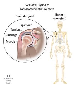
The skeletal system is a complex organ system composed of bones, joints, cartilage, and ligaments. It provides the structural framework for the body and serves several essential physiological roles.
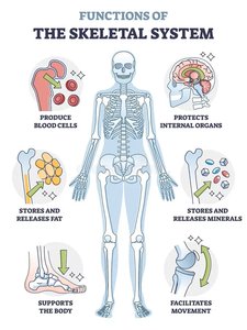
Support: The skeleton supports the body and maintains its shape.
Protection: Bones protect vital organs (e.g., the skull protects the brain, the rib cage protects the heart and lungs).
Leverage: Bones act as levers for muscles, facilitating movement.
Storage: Bones store minerals (such as calcium and phosphate) and lipids (in yellow marrow).
Blood Cell Production: Hematopoiesis occurs in red bone marrow, producing red and white blood cells and platelets.
Classification of Bones
Bone Shapes and Examples
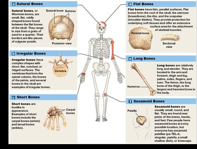
Bones are classified by their shapes, which relate to their functions and locations in the body. Each type of bone may have characteristic markings for muscle attachment, articulation, or passage of nerves and blood vessels.
Sutural Bones: Small, flat, irregular bones found between the flat bones of the skull.
Irregular Bones: Complex shapes (e.g., vertebrae, pelvic bones).
Short Bones: Small and boxy (e.g., carpal bones of the wrist).
Flat Bones: Thin, parallel surfaces (e.g., skull, sternum, ribs, scapulae).
Long Bones: Long and slender (e.g., humerus, femur, radius, ulna, tibia, fibula).
Sesamoid Bones: Small, round, and flat bones found near joints (e.g., patella).
Bone Markings
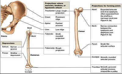
Bone markings are surface features that serve as sites for muscle, tendon, and ligament attachment, or as passages for nerves and blood vessels.
Projections: For attachment or articulation (e.g., trochanter, crest, spine, tubercle, condyle).
Openings and Depressions: For passage of blood vessels and nerves (e.g., foramen, sulcus, fossa).
Structure of a Long Bone
Gross Anatomy
Long bones have a characteristic structure that supports their function in movement and weight-bearing.
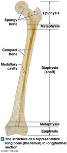
Diaphysis: The shaft, composed of compact bone surrounding a medullary (marrow) cavity.
Epiphysis: The expanded ends, consisting mainly of spongy bone covered by compact bone.
Metaphysis: The narrow region connecting the diaphysis and epiphysis; contains the epiphyseal plate in growing bones.
Composition of Bone
Bone Tissue Components
Bone is a specialized connective tissue composed of cells and an extracellular matrix. The matrix provides strength and flexibility, while the cells are responsible for bone formation, maintenance, and resorption.
Cells: Osteogenic cells, osteoblasts, osteocytes, and osteoclasts.
Matrix: Composed of hydroxyapatite (calcium phosphate crystals) and collagen fibers.
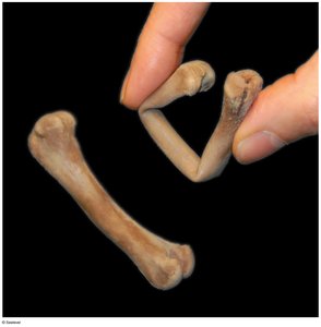
Calcium crystals provide compressive strength but are brittle under twisting or bending forces. Collagen fibers add tensile strength and flexibility.
Bone Cells
There are four main types of bone cells, each with a specific function:
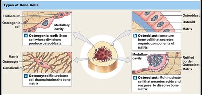
Osteogenic (Osteoprogenitor) Cells: Stem cells that differentiate into osteoblasts; important for fracture repair.
Osteoblasts: Produce new bone matrix (osteoid) and initiate ossification.
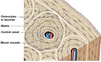
Osteocytes: Mature bone cells that maintain the matrix and participate in repair; reside in lacunae.
Osteoclasts: Large, multinucleated cells that resorb bone matrix (osteolysis).
Compact vs. Spongy Bone
Bone tissue exists in two main forms, each with distinct structure and function:
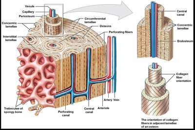
Compact Bone: Dense, organized into osteons (Haversian systems); provides strength and resists stress.
Spongy Bone: Composed of trabeculae; lighter, contains red marrow, and is avascular within the matrix.
Surface Coverings of Bone
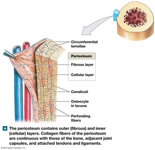
The periosteum is a membrane covering the outer surface of bones, consisting of an outer fibrous layer and an inner cellular layer. It is essential for bone growth, repair, and attachment of tendons and ligaments.
Bone Growth and Development
Ossification and Calcification
Bone formation (ossification or osteogenesis) begins in the embryo and continues until early adulthood. It involves the replacement of other tissues with bone, followed by calcification (deposition of calcium salts).
Endochondral Ossification: Most bones form by replacing hyaline cartilage. Growth occurs in length (interstitial) and width (appositional).
Intramembranous Ossification: Flat bones develop directly from mesenchyme or fibrous connective tissue.
Endochondral Ossification: Steps
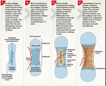
Endochondral ossification is the process by which most bones are formed. The steps include:
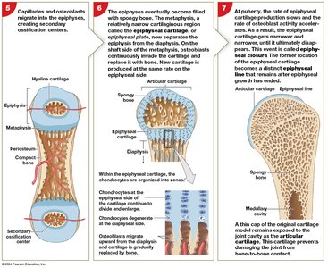
Chondrocytes enlarge and the cartilage matrix calcifies; chondrocytes die.
Blood vessels grow around the cartilage; perichondrium cells become osteoblasts and form a bone collar.
Blood vessels penetrate the cartilage; fibroblasts become osteoblasts and produce spongy bone at the primary ossification center.
Remodeling creates a medullary cavity; bone thickens and cartilage near epiphyses is replaced by bone.
Secondary ossification centers form in the epiphyses.
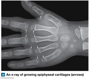
Epiphyses fill with spongy bone; epiphyseal plate separates diaphysis and epiphysis.
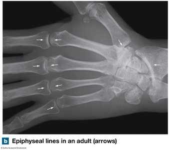
At puberty, ossification outpaces cartilage growth, leading to epiphyseal closure and formation of the epiphyseal line.
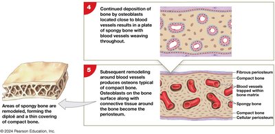
Intramembranous Ossification: Steps
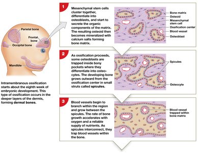
Intramembranous ossification forms flat bones such as the skull, mandible, and clavicles. The steps include:
Mesenchymal cells differentiate into osteoblasts, which secrete osteoid that calcifies to form bone matrix.
Osteoblasts become osteocytes as they are surrounded by bone; bone grows in spicules.
Blood vessels grow between spicules, accelerating bone growth.
Spicules interconnect, trapping blood vessels; a plate of spongy bone forms.
Remodeling produces compact bone on the surface; periosteum forms from osteoblasts and connective tissue.
Bone Remodeling
Continuous Renewal and Homeostasis
Bone remodeling is a lifelong process involving the coordinated activity of osteocytes, osteoblasts, and osteoclasts. It maintains bone strength and mineral homeostasis.
Osteocytes: Remove and replace calcium and protein in the matrix.
Osteoclasts: Resorb bone matrix using acids and enzymes.
Osteoblasts: Deposit new bone matrix.
Homeostatic balance between osteoblast and osteoclast activity is essential. If osteoclasts are more active than osteoblasts, bone mass decreases, potentially leading to osteoporosis.
Exercise stimulates bone remodeling and increases bone density. Lack of weight-bearing activity leads to bone loss and increased fracture risk.
