 Back
The Skeletal System: Structure, Function, and Regional Anatomy CH 8
Study Guide - Smart Notes
 Tailored notes based on your materials, expanded with key definitions, examples, and context.
The Skeletal System
Overview of the Skeleton
The human skeleton provides the structural framework for the body, supports movement, and protects vital organs. It is divided into two main regions: the axial skeleton and the appendicular skeleton.
Axial skeleton: Forms the central supporting axis, including the skull, vertebral column, sternum, ribs, sacrum, and hyoid bone.
Appendicular skeleton: Comprises the pectoral girdle, upper limbs, pelvic girdle, and lower limbs.
Number of bones: The typical adult skeleton contains 206 bones, but this number can vary due to the development of sesamoid bones (e.g., patella) and sutural bones in the skull. At birth, there are about 270 bones, which decrease as some bones fuse during growth.

Anatomical Features of Bones
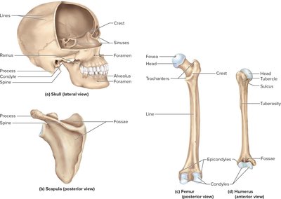
Bones display a variety of markings that serve as sites for muscle attachment, articulation, and passage of nerves and blood vessels. These include ridges, spines, bumps, depressions, canals, foramina, and articular surfaces.
The Skull
General Structure and Cavities
The skull is the most complex part of the skeleton, consisting of 22 bones joined by immovable joints called sutures. It houses several cavities:
Cranial cavity: Encloses the brain.
Orbits: Eye sockets.
Nasal cavity and oral (buccal) cavity
Middle- and inner-ear cavities
Paranasal sinuses: Air-filled spaces in the frontal, sphenoid, ethmoid, and maxillary bones that lighten the skull and add resonance to the voice.
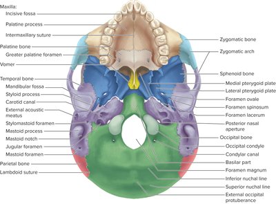
Cranial Bones
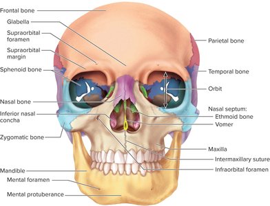
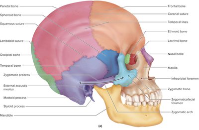
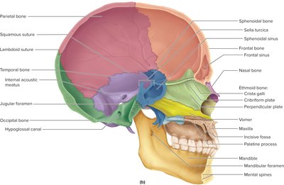
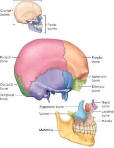
The cranium is divided into cranial bones (which enclose the brain) and facial bones (which shape the face and support cavities). The cranial base is divided into three fossae:
Anterior cranial fossa: Holds the frontal lobes of the brain.
Middle cranial fossa: Holds the temporal lobes.
Posterior cranial fossa: Contains the cerebellum.
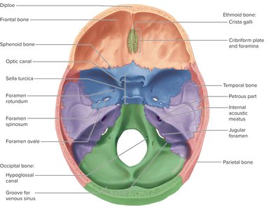
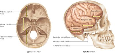
There are eight cranial bones:
1 frontal
2 parietal
2 temporal
1 occipital
1 sphenoid
1 ethmoid
Key Cranial Bones and Features
Frontal bone: Forms the forehead and part of the cranial roof; features include the coronal suture, supraorbital margin, and glabella.
Parietal bones: Form most of the cranial roof and part of its lateral walls; bordered by sagittal, coronal, lambdoid, and squamous sutures.
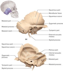
Temporal bones: Form lateral walls and part of the cranial floor; divided into squamous, tympanic, mastoid, and petrous parts.
Occipital bone: Forms the rear and base of the skull; features include the foramen magnum, occipital condyles, and external occipital protuberance.
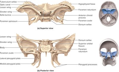
Sphenoid bone: Complex bone with greater and lesser wings, sella turcica (houses the pituitary gland), and several foramina for nerves and vessels.
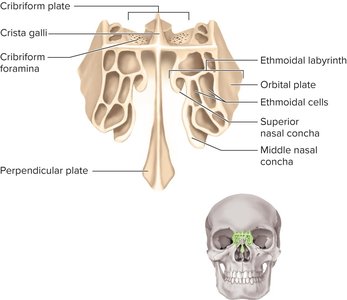
Ethmoid bone: Located between the eyes; forms part of the nasal septum, nasal cavity, and medial wall of the orbit. Features include the cribriform plate, crista galli, and ethmoidal labyrinth.
Facial Bones
There are 14 facial bones, which support the orbital, nasal, and oral cavities, shape the face, and provide attachment for muscles of facial expression and mastication.
2 maxillae
2 palatine
2 zygomatic
2 lacrimal
2 inferior nasal conchae
2 nasal
1 vomer
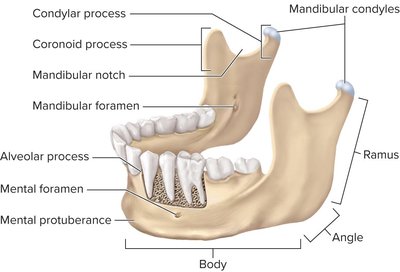
1 mandible
Bones Associated with the Skull
Auditory ossicles: Three small bones in each middle ear (malleus, incus, stapes) involved in hearing.
Hyoid bone: U-shaped bone between the chin and larynx; does not articulate with any other bone and serves as an attachment for muscles of swallowing and speech.
The Skull in Infancy and Childhood
Fontanelles are spaces between unfused cranial bones in infants, allowing for brain growth and passage through the birth canal. The skull approaches adult size by 8 or 9 years of age.
The Vertebral Column and Thoracic Cage
General Features of the Vertebral Column
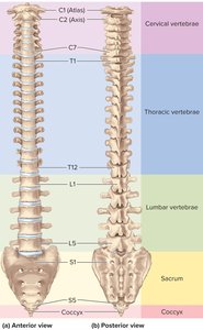
The vertebral column (spine) is a flexible chain of 33 vertebrae separated by intervertebral discs. It supports the skull and trunk, protects the spinal cord, absorbs stress, and provides attachment for limbs and muscles.
7 cervical vertebrae (neck)
12 thoracic vertebrae (chest)
5 lumbar vertebrae (lower back)
5 sacral vertebrae (fused to form the sacrum)
4 coccygeal vertebrae (fused to form the coccyx)
Spinal Curvatures
The adult vertebral column has four curvatures: cervical and lumbar lordosis (concave posteriorly), thoracic and pelvic kyphosis (concave anteriorly). Primary curvatures are present at birth; secondary curvatures develop with head lifting and walking.
General Structure of a Vertebra
Body (centrum): Weight-bearing portion.
Vertebral arch: Formed by pedicles and laminae, enclosing the vertebral foramen.
Processes: Spinous, transverse, and articular processes for muscle and ligament attachment.
Intervertebral foramen: Openings for spinal nerves.
Intervertebral Discs
Discs are pads of fibrocartilage between vertebral bodies, consisting of a gelatinous nucleus pulposus and a tough anulus fibrosus. They bind vertebrae, absorb shock, and allow movement. Herniated discs can compress nerves, causing pain.
Regional Differences in Vertebrae
Cervical vertebrae (C1–C7): Small, with transverse foramina. C1 (atlas) supports the skull; C2 (axis) allows rotation.
Thoracic vertebrae (T1–T12): Larger bodies, costal facets for rib articulation, long downward spinous processes.
Lumbar vertebrae (L1–L5): Thick, stout bodies, blunt spinous processes, resistant to twisting.
Sacrum: Five fused vertebrae forming the posterior pelvic wall.
Coccyx: Four fused vertebrae forming the tailbone.
The Thoracic Cage
The thoracic cage consists of the thoracic vertebrae, sternum, and ribs. It protects the heart and lungs, supports the shoulder girdle, and aids in respiration.
Sternum: Manubrium, body, and xiphoid process.
Ribs: 12 pairs; true ribs (1–7) attach directly to the sternum, false ribs (8–12) do not. Ribs 11 and 12 are floating ribs.
The Pectoral Girdle and Upper Limb
The Pectoral Girdle
The pectoral girdle supports the upper limb and connects it to the axial skeleton. It consists of the clavicle and scapula.
Clavicle: S-shaped bone that braces the shoulder and articulates with the sternum and scapula.
Scapula: Triangular bone with spine, acromion, coracoid process, and glenoid cavity for articulation with the humerus.
The Upper Limb
Humerus: Bone of the arm, articulates with the scapula at the shoulder and with the radius and ulna at the elbow.
Radius and Ulna: Bones of the forearm; radius is lateral, ulna is medial.
Carpals, Metacarpals, Phalanges: Bones of the wrist, palm, and fingers, respectively.
The Pelvic Girdle and Lower Limb
The Pelvic Girdle
The pelvic girdle is a ring of bone composed of two hip bones and the sacrum. It supports the trunk, protects pelvic organs, and anchors the lower limbs.
Hip bone: Formed by the fusion of the ilium, ischium, and pubis.
Pelvic brim, inlet, and outlet: Define the boundaries of the pelvic cavity.
The Lower Limb
Femur: Longest and strongest bone, articulates with the pelvis and tibia.
Patella: Sesamoid bone in the tendon of the quadriceps femoris muscle.
Tibia and Fibula: Bones of the leg; tibia is medial and weight-bearing, fibula is lateral and stabilizes the ankle.
Tarsals, Metatarsals, Phalanges: Bones of the ankle, foot, and toes, respectively.
Arches of the Foot
The foot has three arches (medial longitudinal, lateral longitudinal, and transverse) that absorb stress and provide support. Flat feet (pes planus) can result from weakened ligaments or excessive stress.
