Back
BackThe Skeletal System: Structure, Function, Development, and Pathology
Study Guide - Smart Notes
 Tailored notes based on your materials, expanded with key definitions, examples, and context.
The Skeletal System: Overview
Functions and Tissues of the Skeletal System
The skeletal system is a vital organ system responsible for providing support, protection, and movement to the human body. It is composed of three main types of connective tissues: cartilage, ligaments, and bones.
Support: Provides structural framework for the body.
Protection: Shields vital organs (e.g., skull protects the brain, rib cage protects the heart and lungs).
Movement: Acts as levers for muscles to produce movement.
Blood Cell Formation: Occurs in red bone marrow (hematopoiesis).
Mineral Storage: Stores calcium and phosphate ions.
The three connective tissues are:
Cartilage: Flexible, smooth tissue found in joints, ear, nose, and as a precursor to bone in development.
Ligaments: Dense fibrous connective tissue connecting bone to bone, providing joint stability.
Bones: Hard elements composed of a mineralized matrix and living cells.
Classification and Structure of Bones
Types of Bones
Bones are classified based on their shapes and functions:
Long Bones: Longer than they are wide (e.g., femur, humerus).
Short Bones: Cube-like (e.g., wrist bones).
Flat Bones: Thin and broad (e.g., ribs, sternum).
Irregular Bones: Complex shapes (e.g., vertebrae).
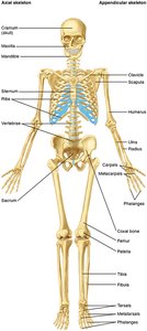
Major Bones of the Human Skeleton
The human skeleton consists of 206 bones, divided intothe axial and appendicular skeletons. Major bones include the skull, vertebral column, ribs, sternum, pelvis, and bones of the limbs.
Axial Skeleton: Skull, vertebral column, rib cage. L a
Appendicular Skeleton: Limbs and girdles (pectoral and pelvic).
Bone Structure
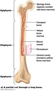
Long Bone Anatomy
Long bones have a distinct structure that supports their function in movement and weight-bearing:
Diaphysis: The shaft, composed mainly of compact bone and containing yellow bone marrow.
Epiphysis: The ends of the bone, containing spongy bone filled with red bone marrow.
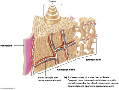
Periosteum: Tough outer membrane containing bone-forming cells (osteocytes).
Compact Bone: Dense outer layer providing strength.
Spongy Bone: Porous inner layer housing marrow.
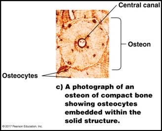
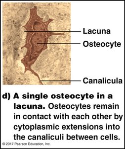
Microscopic Structure of Bone
Bones are organized into cylindrical structures called osteons, which contain concentric rings of bone matrix and osteocytes (mature bone cells) in lacunae. Nutrients are delivered via the central (Haversian) canal, and osteocytes communicate through canaliculi.
Bone Development and Growth
Ossification: From Embryo to Adulthood
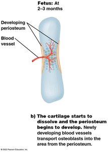
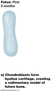
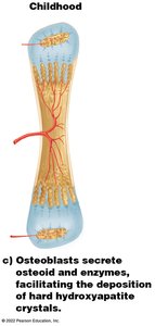
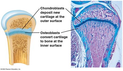
Bone development (ossification) begins in the embryo with the formation of a cartilage model, which is gradually replaced by bone tissue. This process continues through childhood and adolescence as bones grow in length and width.
Chondroblasts: Form hyaline cartilage model in early development.
Osteoblasts: Replace cartilage with bone by secreting osteoid and enzymes for mineralization.
Epiphyseal Plate: Growth plate where new cartilage is produced and converted to bone, allowing lengthening.
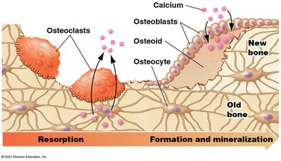
Bone Remodeling and Repair
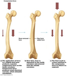
Bone is a dynamic tissue that undergoes continuous remodeling in response to mechanical stress and calcium levels. Osteoclasts resorb bone, while osteoblasts form new bone. This process allows bones to adapt to stress and repair after injury.
Osteoclasts: Break down bone matrix, releasing calcium into the blood.
Osteoblasts: Build new bone matrix.
Mechanical Stress: Increases bone mass and strength in areas of high stress.
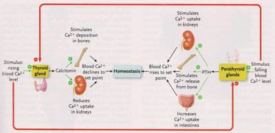
Hormonal Regulation of Bone Growth
Bone remodeling is regulated by hormones to maintain calcium homeostasis:
Calcitonin: Secreted by the thyroid gland when blood calcium is high; stimulates osteoblasts and inhibits osteoclasts, promoting calcium deposition in bone.
Parathyroid Hormone (PTH): Secreted by the parathyroid glands when blood calcium is low; stimulates osteoclasts to release calcium from bone and increases calcium absorption in the kidneys and intestines.
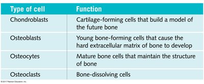
Bone Cells and Their Functions
Type of cell | Function |
|---|---|
Chondroblasts | Cartilage-forming cells that build a model of the future bone |
Osteoblasts | Young bone-forming cells that cause the hard extracellular matrix of bone to develop |
Osteocytes | Mature bone cells that maintain the structure of bone |
Osteoclasts | Bone-dissolving cells |
Pathology of the Skeletal System
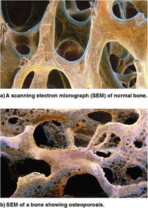
Osteoporosis
Osteoporosis is a condition characterized by decreased bone mass and increased fracture risk, especially in older women due to declining estrogen levels. It results from an imbalance between osteoclast and osteoblast activity.
Symptoms: Brittle bones, hunched posture, difficulty walking, increased fractures (hips, spine).
Prevention: Adequate calcium and vitamin D intake, weight-bearing exercise.
Treatment: Hormone replacement, medications to inhibit osteoclasts, teriparatide (stimulates osteoblasts).
Bone Fractures (Breaks)
Bone fractures are classified by the extent of damage:
Simple: Bone ends do not damage surrounding tissue.
Complete: Bone separates into two pieces, damaging soft tissue.
Compound: Bone ends puncture the skin.
Repair involves hematoma formation, callus formation by chondroblasts, removal of dead tissue by osteoclasts, and new bone formation by osteoblasts.
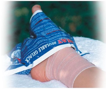
Sprains
Sprains involve stretched or torn ligaments, leading to internal bleeding, bruising, swelling, and pain. Healing is slow due to poor blood supply. Treatment follows the RICE protocol: Rest, Ice, Compression, Elevation.
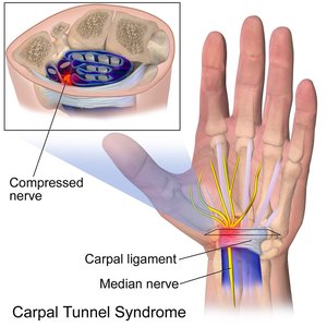
Carpal Tunnel Syndrome
Carpal tunnel syndrome is caused by repetitive use, leading to inflammation of tendons in the wrist and compression of the median nerve. Symptoms include pain, tingling, numbness, and decreased grip strength. Treatments include medications, exercise, and surgery if necessary.
Bursitis and Tendinitis
Bursitis is inflammation of the bursa (fluid-filled sac) near joints, while tendinitis is inflammation of a tendon. Both conditions cause pain, swelling, and restricted movement. Treatment includes ice, heat, rest, and anti-inflammatory medications.
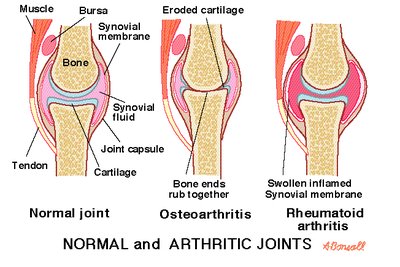
Arthritis
Arthritis is a general term for joint inflammation. The two main types are:
Osteoarthritis: Degeneration of cartilage due to wear and tear, causing pain and inflammation.
Rheumatoid Arthritis: Autoimmune disorder where the immune system attacks joint tissues, leading to deformity and chronic pain.
Treatments include medications, physical therapy, exercise, and sometimes surgery.