 Back
The Skeleton: Structure and Function in Human Anatomy
Study Guide - Smart Notes
 Tailored notes based on your materials, expanded with key definitions, examples, and context.
The Skeleton
Divisions of the Skeleton
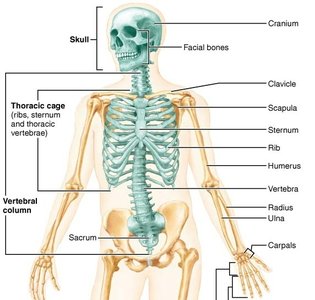
The human skeleton is divided into two main regions: the axial and appendicular skeleton. Each division plays a distinct role in support, protection, and movement.
Axial Skeleton: Includes the skull, vertebral column, and thoracic cage. It forms the long axis of the body and supports the head, neck, and trunk, while protecting the brain, spinal cord, and vital organs of the thorax.
Appendicular Skeleton: Comprises the pectoral girdle and upper limbs, as well as the pelvic girdle and lower limbs. It is responsible for movement and manipulation of the environment.
Axial Skeleton
Skull (Cranium)
The skull is the body's most complex bony structure, housing the brain and forming the framework for the face. It consists of 22 bones: 8 cranial and 14 facial bones.
Cranial Bones: Enclose and protect the brain, provide attachment points for head and neck muscles, and are divided into the vault (calvaria) and base.
Facial Bones: Form the frame for the face, create cavities for sense organs, secure teeth, and provide attachment for facial muscles.
Openings: Approximately 85 named openings allow passage for nerves, blood vessels, and the spinal cord.


Major Cranial Bones
Frontal Bone: Forms the forehead and superior wall of the orbits. Articulates with parietal, ethmoid, sphenoid, lacrimal, nasal, maxillae, and zygomatic bones.
Parietal Bones (2): Form most of the cranial vault. Articulate with frontal (coronal suture), each other (sagittal suture), occipital (lambdoid suture), temporal (squamous suture), and sphenoid (sphenoparietal suture).
Occipital Bone: Forms the posterior wall and base of the cranium. Contains the foramen magnum for passage of the spinal cord and occipital condyles for articulation with the first cervical vertebra.
Temporal Bones (2): Lie inferior to parietal bones. Include squamous, petrous, and tympanic parts, and contain important foramina for nerves and vessels.
Sphenoid: Bat-shaped bone, keystone of the cranium, articulates with all other cranial bones. Contains sella turcica for the pituitary gland and multiple foramina for nerves and vessels.
Ethmoid: Complicated shape, forms most of the bony area between the nasal cavity and orbits. Contains crista galli, cribriform plates, perpendicular plate, and nasal conchae.

Facial Bones
Fourteen bones form the face, most of which are paired. The unpaired bones are the mandible, hyoid, and vomer.
Mandible: Largest and strongest bone of the face, forms the lower jaw and articulates with the temporal bone at the temporomandibular joint (TMJ).
Maxillae (2): Upper jaw bones, keystone of the face, articulate with all other facial bones except the mandible.
Zygoma (2): Form the cheekbones and part of the orbit.
Nasal Bones (2): Form the bridge of the nose.
Lacrimal Bones (2): Form part of the medial orbit and contain the lacrimal fossa for tear drainage.
Palatine Bones (2): Form the posterior hard palate and part of the nasal cavity.
Vomer: Forms part of the nasal septum.
Inferior Nasal Conchae (2): Thin, curved bones projecting into the nasal cavity.
Special Cavities: Orbits
The orbits are formed from seven bones and house the eyes, cushioned by fat and moved by muscles. They contain passageways for nerves and blood vessels.
Vertebral Column
Regions and Curvatures
The vertebral column consists of five major regions and four curvatures, providing support, flexibility, and protection for the spinal cord.
Cervical: 7 vertebrae (C1-C7)
Thoracic: 12 vertebrae (T1-T12)
Lumbar: 5 vertebrae (L1-L5)
Sacrum: 5 fused vertebrae
Coccyx: 3-5 fused vertebrae
Curvatures: Cervical and lumbar (concave posteriorly, secondary), thoracic and sacral (convex posteriorly, primary)
Intervertebral Discs
Intervertebral discs allow movement and act as shock absorbers. They consist of an outer annulus fibrosus and an inner nucleus pulposus.
Annulus Fibrosus: Tough, fibrous tissue
Nucleus Pulposus: Gel-like center
Ligaments and Muscles
Five major ligaments stabilize the vertebral column:
Anterior/Posterior Longitudinal Ligaments: Resist hyperextension/flexion
Ligamentum Flavum: Elastic, flexible, strong
Supraspinous/Interspinous Ligaments: Stabilize spinous processes
Abnormal Spinal Curvatures
Lordosis: Exaggerated lumbar curvature, common in obesity and pregnancy
Kyphosis: Exaggerated thoracic curvature, often due to osteoporosis
Scoliosis: Abnormal lateral bending, often idiopathic or congenital
Herniated Discs
Occurs when the annulus fibrosus tears and the nucleus pulposus protrudes, causing pain and nerve compression.
General Structure of Vertebrae
Body, Vertebral Foramen, Vertebral Arch, Laminae, Pedicles, Intervertebral Foramina
Processes: Spinous, transverse, superior/inferior articular
Special Vertebral Characteristics
C1 (Atlas): No true body, allows nodding
C2 (Axis): Odontoid process, allows rotation
C3-C7: Small, lightweight, greatest range of motion
Thoracic: Heart-shaped bodies, facets for ribs, restricted movement
Lumbar: Large, thick bodies, limited rotation
Sacrum & Coccyx: Fused vertebrae, form pelvic base
Thoracic Cage
Structure and Function
The thoracic cage protects vital organs and supports the shoulder girdles and upper limbs. It consists of the thoracic vertebrae, ribs, and sternum.
Sternum: Long, flat bone with three sections; articulates with clavicles and costal cartilages of ribs 1-7
Ribs: 12 pairs; true ribs (1-7) attach directly to sternum, false ribs (8-12) attach indirectly or not at all, floating ribs (11-12) have no anterior attachment
Appendicular Skeleton
Pectoral Girdle
The pectoral girdle consists of the clavicle and scapula, attaching the upper limbs to the axial skeleton and allowing for great mobility.
Clavicle: S-shaped bone, articulates with sternum and scapula, easily fractured
Scapula: Triangular bone, articulates with humerus at glenoid fossa, provides muscle attachments
Upper Limb
Arm: Humerus
Forearm: Radius (lateral), Ulna (medial)
Hand: Carpals (wrist, 8 bones), Metacarpals (hand, 5 bones), Phalanges (fingers, 14 bones)

Pelvic Girdle
The pelvic girdle attaches to the axial skeleton at the sacroiliac joints and consists of the coxal bones (ilium, ischium, pubis), which fuse in adulthood.
Ilium: Superior, forms hip points
Ischium: Inferior, forms sitz bones
Pubis: Anterior, forms pubic symphysis
Lower Limb
Thigh: Femur (longest, strongest bone), Patella (sesamoid bone)
Leg: Tibia (medial, weight-bearing), Fibula (lateral, stability)
Foot: Tarsals (7 bones), Metatarsals (5 bones), Phalanges (15 bones)
Table: Major Bones of the Human Skeleton
Region | Major Bones | Function |
|---|---|---|
Skull | Frontal, Parietal, Occipital, Temporal, Sphenoid, Ethmoid, Mandible, Maxillae, Zygoma, Nasal, Lacrimal, Palatine, Vomer, Inferior Nasal Conchae | Protection, support, muscle attachment |
Vertebral Column | Cervical, Thoracic, Lumbar, Sacrum, Coccyx | Support, flexibility, protection |
Thoracic Cage | Sternum, Ribs | Protection, support |
Pectoral Girdle | Clavicle, Scapula | Attachment, mobility |
Upper Limb | Humerus, Radius, Ulna, Carpals, Metacarpals, Phalanges | Movement, manipulation |
Pelvic Girdle | Ilium, Ischium, Pubis | Attachment, support |
Lower Limb | Femur, Patella, Tibia, Fibula, Tarsals, Metatarsals, Phalanges | Movement, support |
Example: Carpal Tunnel Syndrome
Carpal tunnel syndrome is caused by inflammation of flexor tendons in the wrist, compressing the median nerve and causing numbness in digits 1-3 and part of 4. Treatment includes wrist bracing.
Example: Medial Tibial Stress Syndrome
Anterior leg pain along the tibia, often due to inflammation from vigorous exercise. Common in athletes; treatment includes rest, ice, NSAIDs, and proper footwear.
Additional info:
All humans have seven cervical vertebrae, regardless of overall neck length.
The segmented structure of the foot must be arched to support weight, with strong ligaments and tendons maintaining the arches.
