 Back
BackThe Skeleton: Structure and Function of the Human Skeletal System
Study Guide - Smart Notes
 Tailored notes based on your materials, expanded with key definitions, examples, and context.
The Skeleton
Overview of the Skeletal System
The human skeleton is a complex framework composed of bones, cartilages, joints, and ligaments. It provides structural support, protects vital organs, and facilitates movement. The skeleton accounts for approximately 20% of body mass and is divided into two major divisions: the axial skeleton and the appendicular skeleton.
Axial skeleton: Forms the longitudinal axis of the body, supporting the head, neck, and trunk, and protecting the brain, spinal cord, and thoracic organs.
Appendicular skeleton: Comprises the bones of the limbs and their girdles, enabling locomotion and manipulation of the environment.


Axial Skeleton
Regions and Functions
The axial skeleton consists of 80 bones divided into three major regions:
Skull
Vertebral column
Thoracic cage
Its primary functions are to form the body's longitudinal axis, support the head, neck, and trunk, and protect the brain, spinal cord, and thoracic organs.
The Skull
The skull is formed by two sets of bones: cranial bones (cranium) and facial bones. Most skull bones are flat and firmly locked together by immovable joints called sutures, except for the mandible.
Cranial bones: Enclose the brain and provide attachment sites for head and neck muscles.
Facial bones: Form the framework of the face, contain cavities for special sense organs, provide openings for air and food passage, secure teeth, and anchor facial muscles for expression.
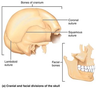
Skull Geography
The cranium is divided into the cranial vault (calvaria) and the cranial base, which forms the inferior aspect of the skull. The base is internally divided into three fossae: anterior, middle, and posterior, which support different regions of the brain.
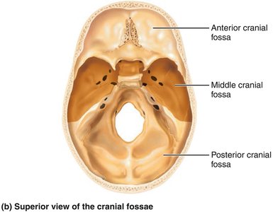
Major Cavities and Openings
Cranial cavity: Houses the brain.
Other cavities: Middle and internal ear cavities, nasal cavity, orbits, and sinuses.
Openings: The skull has 85 named openings (foramina, canals, fissures) for passage of the spinal cord, blood vessels, and cranial nerves.
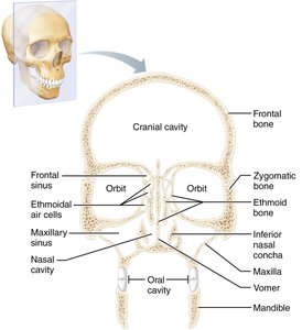
Cranial Bones
Frontal bone
Parietal bones (left and right)
Occipital bone
Temporal bones (left and right)
Sphenoid bone
Ethmoid bone
Facial Bones
Mandible
Maxillary bones (2)
Zygomatic bones (2)
Nasal bones (2)
Lacrimal bones (2)
Palatine bones (2)
Vomer
Inferior nasal conchae (2)
Vertebral Column
General Characteristics
The vertebral column, or spine, extends from the skull to the pelvis and consists of 26 irregular bones called vertebrae, organized into five major regions:
Cervical: 7 vertebrae
Thoracic: 12 vertebrae
Lumbar: 5 vertebrae
Sacrum: 1 bone (fusion of several vertebrae)
Coccyx: 1 bone (fusion of several vertebrae)
The vertebral column transmits the weight of the trunk to the lower limbs, surrounds and protects the spinal cord, and provides attachment points for ribs and muscles.
Curvatures
Cervical and lumbar curvatures: Concave posteriorly
Thoracic and sacral curvatures: Convex posteriorly
These curvatures increase the resilience and flexibility of the spine.
Abnormal Spinal Curvatures
Scoliosis: Abnormal lateral rotation of the spine, often in the thoracic region.
Kyphosis (hunchback): Abnormal dorsal thoracic curvature.
Lordosis (swayback): Accentuated lumbar curvature.
Ligaments and Intervertebral Discs
Ligaments: Support the vertebral column and prevent excessive movement.
Intervertebral discs: Cushionlike pads between vertebrae, acting as shock absorbers. Each disc has a gelatinous nucleus pulposus and a tough anulus fibrosus.
General Structure of Vertebrae
Body: Weight-bearing region
Vertebral arch: Formed by pedicles and laminae
Processes: Spinous, transverse, superior and inferior articular processes
Vertebral foramen: Encloses the spinal cord
Regional Vertebral Characteristics
Characteristic | Cervical | Thoracic | Lumbar |
|---|---|---|---|
Body | Small, oval | Heart-shaped, bears costal facets | Massive, kidney-shaped |
Spinous process | Short, bifid (except C7) | Long, sharp, projects inferiorly | Short, blunt, rectangular |
Vertebral foramen | Triangular, large | Circular | Triangular |
Transverse processes | Contain foramina | Bear facets for ribs | Thin and tapered |
Movements allowed | Flexion, extension, rotation | Rotation, limited flexion/extension | Flexion, extension, some lateral flexion |
Thoracic Cage
Structure and Function
The thoracic cage is composed of the thoracic vertebrae, ribs, sternum, and costal cartilages. It protects vital organs, supports the shoulder girdles and upper limbs, and provides attachment sites for muscles.
Sternum: Consists of the manubrium, body, and xiphoid process.
Ribs: 12 pairs; true ribs (1–7) attach directly to the sternum, false ribs (8–10) attach indirectly, and floating ribs (11–12) have no anterior attachment.
Appendicular Skeleton
Pectoral Girdle
The pectoral (shoulder) girdle consists of the clavicles and scapulae, attaching the upper limbs to the axial skeleton and providing mobility and flexibility.
Clavicle: S-shaped bone articulating with the sternum and scapula.
Scapula: Triangular bone with borders, angles, and several important processes (spine, acromion, coracoid process).
Upper Limb
Arm: Humerus
Forearm: Radius and ulna
Hand: Carpals (wrist), metacarpals (palm), phalanges (fingers)
Pelvic Girdle
The pelvic (hip) girdle is formed by two hip bones and the sacrum, attaching the lower limbs to the axial skeleton and supporting pelvic organs. The coxal bone is formed by the fusion of the ilium, ischium, and pubis.
Lower Limb
Thigh: Femur (largest bone in the body)
Leg: Tibia (weight-bearing) and fibula
Foot: Tarsals, metatarsals, phalanges
Developmental Aspects of the Skeleton
Infant and Childhood Development
Infant skull has more bones than adult skull; bones are connected by fontanelles to allow for growth and birth.
Primary spinal curvatures are present at birth; secondary curvatures develop as the child grows.
Aging and the Skeleton
Intervertebral discs lose hydration and elasticity with age, increasing the risk of herniation.
Costal cartilages ossify, and bone mass decreases, increasing fracture risk.
