 Back
The Skeleton: Structure, Function, and Clinical Relevance
Study Guide - Smart Notes
 Tailored notes based on your materials, expanded with key definitions, examples, and context.
The Skeleton
Introduction
The human skeleton is a complex framework of bones that provides structural support, protection for vital organs, and facilitates movement. Understanding its anatomy is essential for anticipating clinical issues, such as those affecting labor and delivery, and for comprehending the body's overall function.
Axial Skeleton
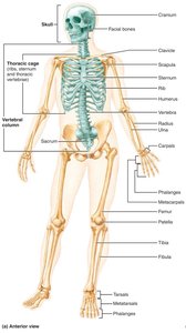
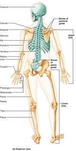
The axial skeleton consists of 80 bones divided into three major regions: the skull, vertebral column, and thoracic cage. Its primary functions are to form the longitudinal axis of the body, support the head, neck, and trunk, and protect the brain, spinal cord, and thoracic organs.
Skull: Protects the brain and forms the structure of the face.
Vertebral column: Supports the trunk and protects the spinal cord.
Thoracic cage: Protects the heart and lungs.
The Skull
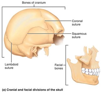
The skull is composed of two main parts: the cranium and the facial bones. The cranium consists of eight bones that enclose the brain, while the facial skeleton is made up of 14 bones, most of which are paired.
Cranial bones: Frontal, parietal (2), occipital, temporal (2), sphenoid, ethmoid.
Facial bones: Mandible, maxillary (2), zygomatic (2), nasal (2), lacrimal (2), palatine (2), vomer, inferior nasal conchae (2).
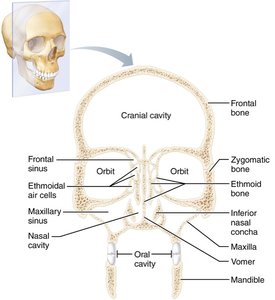
Major Cavities of the Skull
The skull contains several cavities, including the cranial cavity, orbits, nasal cavity, oral cavity, and paranasal sinuses. These cavities house and protect sensory organs and provide passageways for air and food.
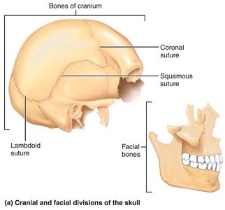
Cranial Sutures
Sutures are immovable joints that connect the bones of the skull. The main sutures include:
Coronal suture: Between parietal bones and frontal bone.
Sagittal suture: Between right and left parietal bones.
Lambdoid suture: Between parietal bones and occipital bone.
Squamous suture: Between parietal and temporal bones.
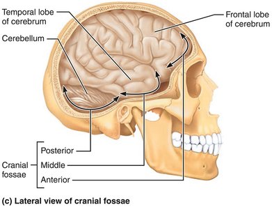
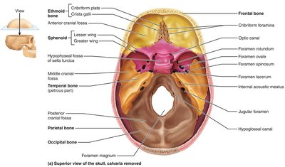
Base of the Cranial Cavity
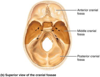
The base of the cranial cavity is divided into three fossae: anterior, middle, and posterior. These depressions accommodate different parts of the brain.
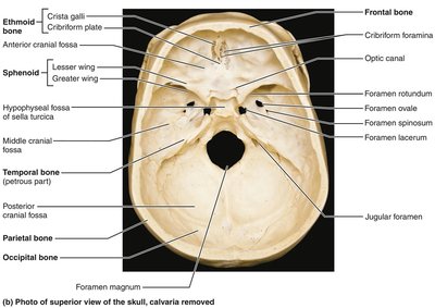
Clinical Relevance: Mastoiditis
The mastoid process contains air cells that can become infected, especially following middle ear infections. Because these cells are separated from the brain by a thin bony plate, infection can potentially spread to the brain.
Facial Bones
The facial bones form the structure of the face, house the teeth, and provide attachment points for muscles involved in facial expression and mastication.
Mandible: Lower jawbone, only movable bone of the skull.
Maxilla: Upper jawbone, forms part of the hard palate.
Zygomatic bones: Cheekbones.
Nasal bones: Bridge of the nose.
Lacrimal bones: Medial wall of the orbit.
Palatine bones: Posterior part of the hard palate.
Vomer: Forms part of the nasal septum.
Inferior nasal conchae: Form part of the lateral walls of the nasal cavity.
The Vertebral Column
The vertebral column, or spine, is a flexible structure composed of 26 vertebrae in five regions. It transmits the weight of the trunk to the lower limbs, protects the spinal cord, and provides attachment points for ribs and muscles.
Cervical: 7 vertebrae (C1–C7)
Thoracic: 12 vertebrae (T1–T12)
Lumbar: 5 vertebrae (L1–L5)
Sacrum: 1 bone (5 fused vertebrae)
Coccyx: 1 bone (4 fused vertebrae)
Intervertebral Discs
Intervertebral discs are cushion-like pads between vertebrae that act as shock absorbers. They consist of:
Nucleus pulposus: Inner gelatinous core providing elasticity and compressibility.
Anulus fibrosus: Outer collar of collagen and fibrocartilage limiting expansion.
Abnormal Spinal Curvatures
Abnormal curvatures can be congenital or acquired:
Scoliosis: Lateral curvature, often in thoracic region.
Kyphosis: Dorsal thoracic curvature (hunchback).
Lordosis: Lumbar curvature (swayback).
Regional Vertebral Characteristics
Each region of the vertebral column has unique features:
Characteristic | Cervical (3–7) | Thoracic | Lumbar |
|---|---|---|---|
Body | Small, oval, wide | Heart-shaped, bears costal facets | Massive, kidney-shaped |
Spinous process | Short, bifid (except C7) | Long, sharp, projects inferiorly | Short, blunt, rectangular |
Vertebral foramen | Triangular, large | Circular | Triangular |
Transverse processes | Contain foramina | Bear facets for ribs | Thin and tapered |
Movements allowed | Flexion, extension, lateral flexion, rotation | Rotation, limited flexion/extension | Flexion, extension, some lateral flexion |
Thoracic Cage
The thoracic cage is composed of the thoracic vertebrae, ribs, sternum, and costal cartilages. It protects vital organs, supports the shoulder girdles and upper limbs, and provides attachment sites for muscles.
Sternum: Manubrium, body, xiphoid process.
Ribs: 12 pairs, classified as true (1–7), false (8–12), and floating (11–12).
Appendicular Skeleton
The appendicular skeleton consists of the bones of the limbs and their girdles. The pectoral girdle attaches the upper limbs to the trunk, while the pelvic girdle attaches the lower limbs.
Pectoral girdle: Clavicle and scapula.
Pelvic girdle: Hip bones (coxal bones) and sacrum.
Upper Limb
Each upper limb contains 30 bones:
Arm: Humerus
Forearm: Radius and ulna
Hand: Carpals (8), metacarpals (5), phalanges (14)
Lower Limb
The lower limb carries the body's weight and is subjected to significant forces. It consists of:
Thigh: Femur
Leg: Tibia and fibula
Foot: Tarsals (7), metatarsals (5), phalanges (14)
Developmental Aspects of the Skeleton
Infant skulls have more bones than adult skulls, with unfused mandible and frontal bones connected by fontanelles. As children grow, the cranium and facial bones change in proportion, and the spine develops secondary curvatures.
Fontanelles: Anterior, posterior, mastoid, sphenoidal.
Primary curvatures: Thoracic and sacral (convex at birth).
Secondary curvatures: Cervical and lumbar (develop with age).
Clinical Relevance
Mastoiditis: Infection of mastoid air cells, risk of spreading to brain.
Herniated discs: Rupture of anulus fibrosus, protrusion of nucleus pulposus.
Colles' fracture: Break in distal radius, common in falls.
Carpal tunnel syndrome: Compression of median nerve in wrist.
Bimalleolar fracture: Break in distal tibia and fibula, common sports injury.
Flat foot (pes planus): Loss of arch, congenital or acquired.
Cleft palate: Failure of medial fusion of palate, affects feeding and breathing.
Developmental dysplasia of the hip (DDH): Dislocation or poor development of hip joint in newborns.
Comparison of Male and Female Pelves
Characteristic | Female | Male |
|---|---|---|
General structure | Tilted forward, adapted for childbearing | Tilted less, adapted for support |
Bone thickness | Lighter, thinner, smoother | Heavier, thicker, more prominent markings |
Acetabula | Smaller, farther apart | Larger, closer together |
Pubic arch | Broader (80–90°) | More acute (50–60°) |
Sacrum | Wider, shorter | Narrow, longer |
Coccyx | More movable, projects inferiorly | Less movable, projects anteriorly |
Pelvic inlet | Wider, oval | Narrow, heart-shaped |
Pelvic outlet | Wider, ischial spines farther apart | Narrower, ischial spines closer together |
