 Back
The Skeleton: Structure, Function, and Regional Anatomy
Study Guide - Smart Notes
 Tailored notes based on your materials, expanded with key definitions, examples, and context.
The Skeleton
Overview of the Skeletal System
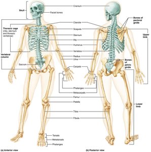
The skeletal system is a complex framework of bones, cartilages, joints, and ligaments that provides structural support, protection, and movement for the human body. It accounts for approximately 20% of body mass and is divided into two major divisions: the axial skeleton and the appendicular skeleton.
Axial skeleton: Composed of 80 bones, including the skull, vertebral column, and rib cage. It forms the longitudinal axis of the body, supports the head, neck, and trunk, and protects the brain, spinal cord, and thoracic organs.
Appendicular skeleton: Consists of 126 bones, including the limbs and girdles, facilitating locomotion and manipulation of the environment.
The Skull
General Structure and Function
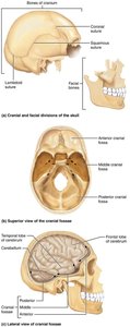
The skull is the most complex bony structure in the body, formed by two sets of bones: cranial bones and facial bones. Most skull bones are flat and joined by immovable joints called sutures, which have a serrated, saw-tooth appearance.
Cranial bones (cranium): Enclose the brain and provide attachment sites for head and neck muscles.
Facial bones: Form the framework of the face, house special sense organs, provide openings for air and food, secure teeth, and anchor facial muscles for expression.
Cranial Bones
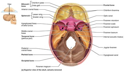
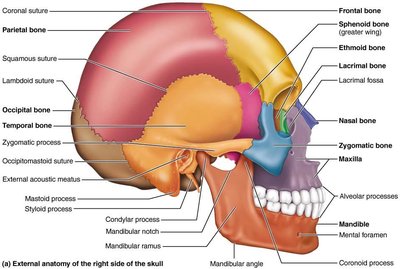
The cranium is comprised of eight bones: frontal, parietal (2), occipital, temporal (2), sphenoid, and ethmoid. These bones protect the brain and form the cranial cavity.
Frontal bone: Forms the forehead and anterior cranium; contains the supraorbital foramen and frontal sinuses.
Parietal bones: Form the superior and lateral aspects of the skull; joined by coronal, sagittal, lambdoid, and squamous sutures.
Occipital bone: Forms the posterior skull and cranial fossa; contains the foramen magnum and occipital condyles.
Temporal bones: Form the inferolateral skull and cranial base; include mastoid and styloid processes, zygomatic process, and external acoustic meatus.
Sphenoid bone: Keystone bone that articulates with all other cranial bones; contains the sella turcica and sphenoidal sinuses.
Ethmoid bone: Deepest skull bone; forms the roof of the nasal cavity and part of the nasal septum; contains cribriform plates, crista galli, and ethmoidal air cells.
Facial Bones
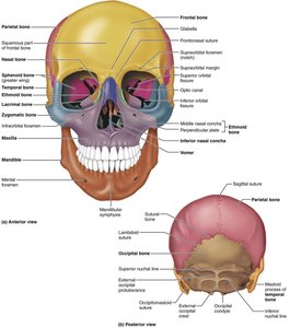
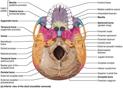
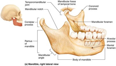
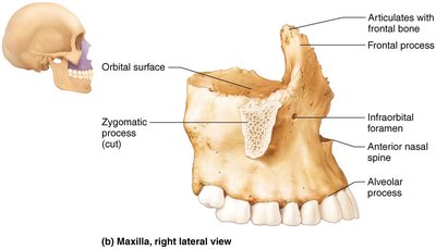
The facial skeleton consists of 14 bones, including the mandible, maxillae, zygomatic, nasal, lacrimal, palatine, vomer, and inferior nasal conchae. These bones shape the face, form cavities for sense organs, and provide passageways for air and food.
Mandible: The largest and strongest facial bone; forms the lower jaw and articulates with the temporal bone at the temporomandibular joint.
Maxillae: Fused medially to form the upper jaw; contain alveolar processes for upper teeth and maxillary sinuses.
Zygomatic bones: Form the cheekbones and part of the orbits.
Nasal bones: Form the bridge of the nose.
Lacrimal bones: Form part of the medial wall of the orbits; contain the lacrimal fossa for tear drainage.
Palatine bones: Form the posterior part of the hard palate and part of the nasal cavity and orbit.
Vomer: Forms part of the nasal septum.
Inferior nasal conchae: Form part of the lateral walls of the nasal cavity.
The Hyoid Bone
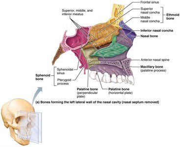
The hyoid bone is unique in that it does not articulate directly with any other bone. Located in the anterior neck, it serves as a movable base for the tongue and an attachment site for muscles involved in swallowing and speech.
Special Features: Orbits, Nasal Cavity, and Paranasal Sinuses
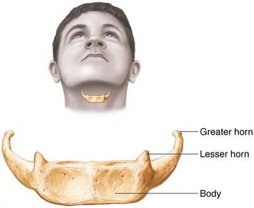
Orbits: Bony cavities that house the eyes and associated structures, formed by seven bones (frontal, sphenoid, zygomatic, maxilla, palatine, lacrimal, ethmoid).
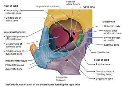
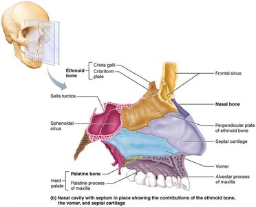
Nasal cavity: Formed by parts of several bones; contains conchae that increase air turbulence and meatuses for airflow regulation.
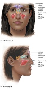
Paranasal sinuses: Air-filled spaces in the frontal, sphenoid, ethmoid, and maxillary bones; function to warm, humidify air, lighten the skull, and enhance voice resonance.
The Vertebral Column
General Characteristics
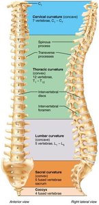
The vertebral column, or spine, extends from the skull to the pelvis and consists of 26 irregular bones in adults. It transmits the weight of the trunk to the lower limbs, protects the spinal cord, and provides attachment points for ribs and muscles. The column is divided into five regions:
Cervical (7 vertebrae): Neck region
Thoracic (12 vertebrae): Upper back, articulates with ribs
Lumbar (5 vertebrae): Lower back, bears most weight
Sacrum (1 bone): Formed from fusion of five vertebrae
Coccyx (1 bone): Formed from fusion of three to five vertebrae
There are four main curvatures: cervical and lumbar (concave posteriorly, secondary), thoracic and sacral (convex posteriorly, primary).
Intervertebral Discs
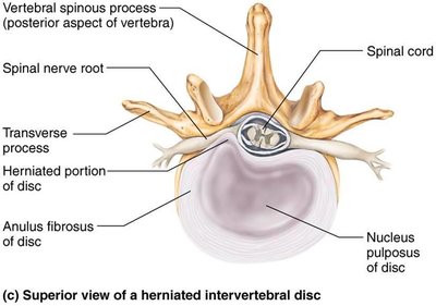
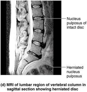
Intervertebral discs are cushion-like pads between vertebrae that act as shock absorbers. Each disc consists of:
Nucleus pulposus: Inner gelatinous core providing elasticity and compressibility.
Anulus fibrosus: Outer collar of collagen and fibrocartilage that limits expansion of the nucleus pulposus.
Abnormal Spinal Curvatures
Scoliosis: Abnormal lateral curvature, often in the thoracic region; may cause breathing difficulties.
Kyphosis: Exaggerated dorsal thoracic curvature (hunchback); common in osteoporosis.
Lordosis: Accentuated lumbar curvature (swayback); seen in pregnancy or obesity.
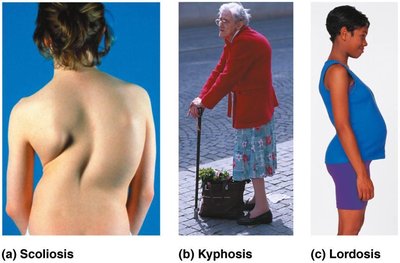
General Structure of Vertebrae
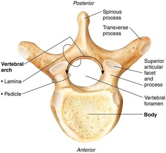
All vertebrae share a common structure:
Body: Anterior, weight-bearing region
Vertebral arch: Formed by pedicles and laminae
Vertebral foramen: Canal for the spinal cord
Processes: Spinous, transverse, superior and inferior articular processes
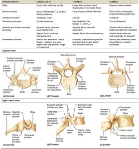
Regional Vertebral Characteristics
Characteristic | Cervical (C3–C7) | Thoracic | Lumbar |
|---|---|---|---|
Body | Small, oval, wide side to side | Larger than cervical; heart-shaped | Massive, kidney-shaped |
Spinous process | Short; bifid (except C7) | Long; sharp; projects inferiorly | Short; blunt; projects directly posteriorly |
Vertebral foramen | Triangular | Circular | Triangular |
Transverse processes | Contain foramina | Bear facets for ribs (except T11, T12) | Thin and tapered |
Movements allowed | Flexion, extension, lateral flexion, rotation | Rotation; limited flexion/extension | Flexion and extension; rotation prevented |
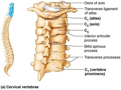
Special Cervical Vertebrae: Atlas (C1) and Axis (C2)
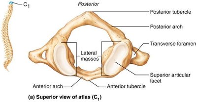
Atlas (C1): No body or spinous process; supports the skull and allows nodding "yes" motion.
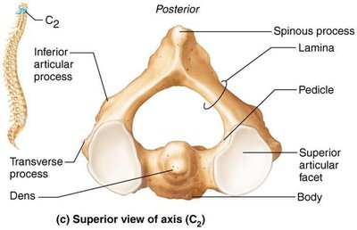
Axis (C2): Has a dens (odontoid process) that acts as a pivot for rotation, allowing the "no" motion.
Thoracic and Lumbar Vertebrae
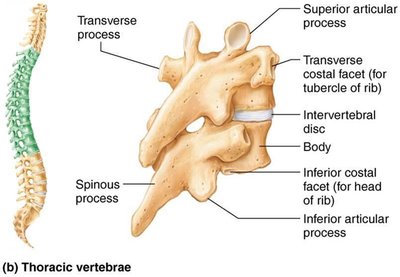
Thoracic vertebrae (T1–T12): Articulate with ribs; have heart-shaped bodies and long, downward-pointing spinous processes.
Lumbar vertebrae (L1–L5): Largest vertebrae; support most body weight; have short, thick processes and prevent rotation.
Sacrum and Coccyx
Sacrum: Triangular bone formed from five fused vertebrae; articulates with the hip bones at the sacroiliac joints.
Coccyx: Tailbone formed from three to five fused vertebrae; vestigial structure with little function.
Summary Table: Major Bones of the Axial Skeleton
Region | Main Bones | Key Functions |
|---|---|---|
Skull | Frontal, parietal, occipital, temporal, sphenoid, ethmoid, mandible, maxilla, zygomatic, nasal, lacrimal, palatine, vomer, inferior nasal conchae | Protects brain, forms face, houses sense organs |
Vertebral Column | Cervical, thoracic, lumbar vertebrae, sacrum, coccyx | Supports trunk, protects spinal cord, attachment for ribs/muscles |
Thoracic Cage | Sternum, ribs, costal cartilages, thoracic vertebrae | Protects thoracic organs, supports shoulder girdles |
Additional info: The above notes provide a comprehensive overview of the skeletal system, focusing on the axial skeleton and its major components, as outlined in Chapter 7 of a standard anatomy and physiology textbook. The included images are directly relevant to the anatomical structures discussed and reinforce the textual explanations.
