 Back
BackThe Skull: Structure, Bones, and Cavities
Study Guide - Smart Notes
 Tailored notes based on your materials, expanded with key definitions, examples, and context.
Module 7.2: The Skull
Overview of Skull Structure
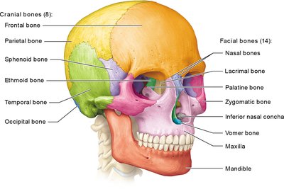
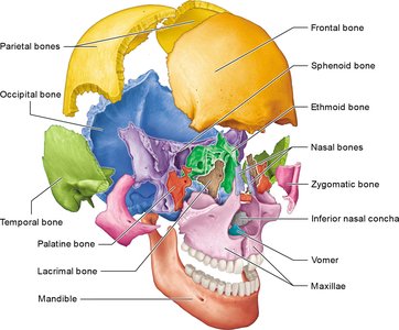
The skull is the most complex bony structure in the human body, consisting of 22 bones (excluding the auditory ossicles). It is divided into two main groups: cranial bones, which encase the brain, and facial bones, which form the framework of the face. These bones also contribute to the protection and housing of special sensory organs.
Cranial bones (8): Frontal, parietal (2), temporal (2), occipital, sphenoid, ethmoid
Facial bones (14): Maxilla (2), zygomatic (2), nasal (2), lacrimal (2), palatine (2), inferior nasal concha (2), vomer, mandible
All skull bones except the mandible are joined by immovable joints called sutures.
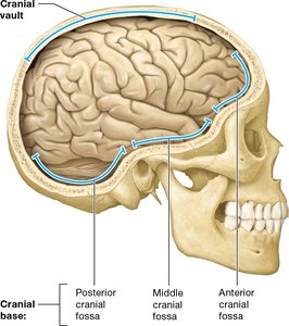
Cranial Cavity and Skull Regions
The cranial bones form the cranial cavity, which houses and protects the brain. The cranial cavity is divided into the cranial vault (calvaria) and the cranial base. The cranial base is further subdivided into three fossae: anterior, middle, and posterior, each accommodating different regions of the brain.
Cranial vault: Superior portion, forms the "roof" of the cranial cavity.
Cranial base: Inferior portion, forms the "floor" of the cranial cavity.
Cranial fossae: Depressions in the cranial base for brain regions.
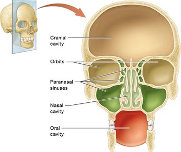
Major Cavities of the Skull
Besides the cranial cavity, the skull contains several smaller cavities that house sensory organs and other structures:
Orbits: Contain the eyeballs and associated structures.
Nasal cavity: Houses sensory receptors for smell and is part of the respiratory tract.
Oral cavity: Surrounds the teeth and tongue, forming the entry to the digestive tract.
Paranasal sinuses: Air-filled spaces in certain skull bones that lighten the skull and enhance voice resonance.
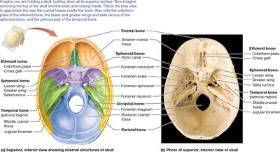
Internal Structure of the Skull
The internal view of the skull reveals the arrangement of cranial fossae and the passageways for nerves and blood vessels. The superior view shows the anterior, middle, and posterior cranial fossae, each formed by different cranial bones.
Anterior cranial fossa: Houses the frontal lobes of the brain.
Middle cranial fossa: Accommodates the temporal lobes.
Posterior cranial fossa: Contains the cerebellum.
Disarticulated Skull
Understanding the skull's complexity is aided by viewing it as a disarticulated structure, where each bone is separated. This "exploded" view helps visualize how the bones fit together, especially complex ones like the ethmoid and sphenoid.
Cavities of the Skull
The Orbit
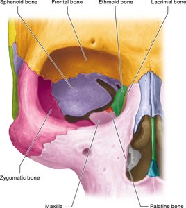
The orbit is a bony cavity that houses the eyeball, associated muscles, nerves, blood vessels, and the lacrimal gland. It is formed by seven bones: frontal, maxilla, zygomatic, sphenoid, ethmoid, lacrimal, and palatine. The complexity of the orbit makes injuries challenging to treat.
Nasal Cavity and Paranasal Sinuses
The nasal cavity is the entryway to the respiratory tract, formed by several bones and lined with mucous membranes. The paranasal sinuses are air-filled spaces in the frontal, sphenoid, ethmoid, and maxillary bones, connected to the nasal cavity. They function to filter, warm, and humidify air, lighten the skull, and enhance voice resonance.
Posterior wall: Sphenoid body and pterygoid processes
Lateral walls: Ethmoid, palatine, inferior nasal conchae, maxilla
Roof: Cribriform plate of ethmoid
Floor: Hard palate (palatine bones and maxillae)
Nasal septum: Hyaline cartilage (anterior), perpendicular plate of ethmoid and vomer (posterior)
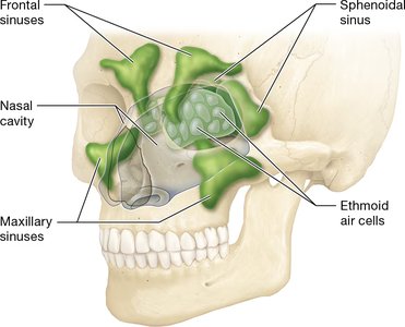
Clinical note: Infections can spread easily between the nasal cavity and sinuses, leading to sinusitis.
Oral Cavity
The oral cavity contains the teeth, tongue, and salivary glands. Its roof is the hard palate, and its walls are formed by the maxillae and mandible. The floor and posterior wall are made of soft tissues.
Fetal and Infant Skull
Fontanels and Skull Flexibility
Infant skulls have "soft spots" called fontanels, which are membranous areas where ossification is incomplete. These allow for flexibility during birth and accommodate brain growth. Major fontanels include the anterior, posterior, sphenoid, and mastoid fontanels. Sutures are also unfused in infants, providing additional flexibility.
Clinical note: The degree of suture fusion can help estimate age in forensic investigations.
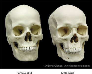
Sex Differences in the Skull
Male and female skulls have distinct features. Males typically have a sloped forehead, prominent supraorbital ridge, a mandibular angle near 90 degrees, and a larger mastoid process. Females have a straighter forehead, less prominent ridge, a wider mandibular angle, and a smaller mastoid process.
The Hyoid Bone
Structure and Function
The hyoid bone is a small, C-shaped bone in the superior neck. It does not articulate with any other bone but is suspended by muscles and ligaments. It serves as an attachment point for muscles involved in swallowing and speech. In forensic science, a fractured hyoid bone may indicate strangulation.
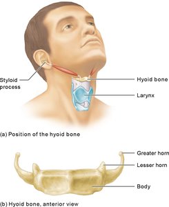
Table: Major Cranial and Facial Bones
Bone | Location | Main Function(s) |
|---|---|---|
Frontal | Forehead, roof of orbits | Protects brain, forms upper face |
Parietal | Superior and lateral skull | Protects brain |
Temporal | Inferior lateral skull | Protects brain, houses ear structures |
Occipital | Posterior skull | Protects brain, contains foramen magnum |
Sphenoid | Base of skull | Supports brain, forms part of orbits |
Ethmoid | Medial orbit, nasal cavity | Supports nasal cavity, forms part of orbits |
Maxilla | Upper jaw | Holds upper teeth, forms part of orbit and palate |
Mandible | Lower jaw | Holds lower teeth, only movable skull bone |
Zygomatic | Cheek | Forms cheekbone, part of orbit |
Nasal | Bridge of nose | Supports nose cartilage |
Lacrimal | Medial orbit | Houses lacrimal sac |
Palatine | Posterior hard palate | Forms part of palate and nasal cavity |
Inferior nasal concha | Lateral nasal cavity | Filters and humidifies air |
Vomer | Nasal septum | Divides nasal cavity |
