 Back
BackThe Skull: Structure, Bones, and Key Features
Study Guide - Smart Notes
 Tailored notes based on your materials, expanded with key definitions, examples, and context.
The Skull: Structure and Overview
Introduction to the Skull
The skull is the most complex bony structure in the human body, composed of cranial and facial bones. It serves to protect the brain, support sensory structures, and provide attachment points for muscles involved in facial expression and mastication.
Cranial bones (cranium): Enclose and protect the brain, provide muscle attachment sites.
Facial bones: Form the framework of the face, house cavities for special senses, provide openings for air and food, secure teeth, and anchor facial muscles.
Most skull bones are flat bones joined by immovable sutures, except the mandible, which is connected by a movable joint.
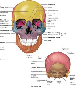
Major Sutures of the Skull
Coronal suture: Between frontal and parietal bones
Sagittal suture: Between right and left parietal bones
Lambdoid suture: Between parietal and occipital bones
Squamous suture: Between parietal and temporal bones
Geography of the Skull
Cranial Vault and Base
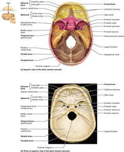
The cranium is divided into the cranial vault (calvaria) and the cranial base:
Cranial vault: Forms the superior, lateral, and posterior aspects of the skull, including the forehead.
Cranial base: Forms the inferior aspect, divided into anterior, middle, and posterior cranial fossae, which cradle the brain.
The cranial cavity is the space enclosed by the vault and base, housing the brain.
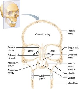
Other Cavities and Openings
Additional cavities: Middle and internal ear cavities, nasal cavity, orbits (eye sockets).
Several bones contain paranasal sinuses that lighten the skull and enhance resonance of the voice.
Numerous foramina, canals, and fissures allow passage of nerves and blood vessels.
Cranial Bones
Overview of Cranial Bones
The cranium consists of eight bones: paired parietal and temporal bones, and unpaired frontal, occipital, sphenoid, and ethmoid bones. These bones form a protective 'helmet' for the brain.
Frontal Bone
Forms the forehead, superior wall of the orbits, and most of the anterior cranial fossa.
Features: Supraorbital margins (under eyebrows), supraorbital foramen (passage for artery and nerve), glabella (smooth area between orbits), and frontal sinuses.
Parietal Bones and Major Sutures
Form most of the superior and lateral aspects of the skull.
Articulate with other cranial bones at the four major sutures: coronal, sagittal, lambdoid, and squamous.
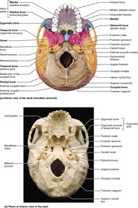
Occipital Bone
Forms the posterior wall and base of the skull.
Features: Foramen magnum (passage for spinal cord), occipital condyles (articulate with first vertebra), external occipital protuberance, and nuchal lines (muscle attachment).
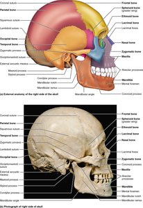
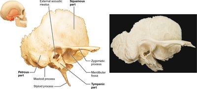
Temporal Bones
Form the inferolateral aspects of the skull and part of the cranial base.
Three major parts: Squamous (zygomatic process, mandibular fossa), tympanic (external acoustic meatus), and petrous (houses middle and inner ear, mastoid and styloid processes).
Important foramina: Jugular foramen, carotid canal, foramen lacerum, internal acoustic meatus.
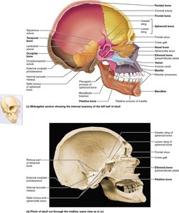
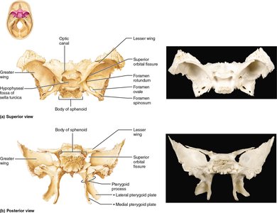
Sphenoid Bone
Keystone bone of the cranium, articulates with all other cranial bones.
Features: Body, greater and lesser wings, pterygoid processes, sella turcica (houses pituitary gland), sphenoidal sinuses.
Openings: Optic canal, superior orbital fissure, foramen rotundum, foramen ovale, foramen spinosum.
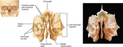
Ethmoid Bone
Most deeply situated cranial bone, forms the bony area between the nasal cavity and orbits.
Features: Cribriform plates (with cribriform foramina for olfactory nerves), crista galli (attachment for brain covering), perpendicular plate (part of nasal septum), ethmoidal air cells, superior and middle nasal conchae.
Sutural Bones
Tiny, irregular bones found within sutures, most often in the lambdoid suture. Their significance is unknown.
Facial Bones
Overview of Facial Bones
The facial skeleton consists of 14 bones: mandible, vomer (unpaired), maxillae, zygomatics, nasals, lacrimals, palatines, and inferior nasal conchae (paired). The maxillae are the keystone bones of the face.
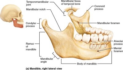
Mandible
Largest, strongest facial bone; forms the lower jaw.
Features: Body, rami, mandibular angle, coronoid process, condylar process (forms temporomandibular joint), alveolar process (anchors teeth), mandibular and mental foramina.
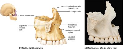
Maxillary Bones (Maxillae)
Form the upper jaw and central portion of the facial skeleton; all facial bones except the mandible articulate with the maxillae.
Features: Alveolar processes (anchor upper teeth), palatine processes (form anterior hard palate), maxillary sinuses, infraorbital foramen.
Zygomatic Bones
Form the cheekbones and part of the inferolateral margins of the orbits.
Nasal Bones
Fused medially to form the bridge of the nose.
Lacrimal Bones
Contribute to the medial walls of each orbit; contain a groove for the lacrimal sac (tear drainage).
Palatine Bones
L-shaped bones forming the posterior part of the hard palate and part of the nasal cavity and orbits.
Vomer
Plow-shaped bone forming part of the nasal septum.
Inferior Nasal Conchae
Thin, curved bones projecting into the nasal cavity, forming part of its lateral walls.
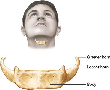
The Hyoid Bone
Structure and Function
Located in the anterior neck, inferior to the mandible.
Unique: does not articulate directly with any other bone; anchored by ligaments to the styloid processes of the temporal bones.
Acts as a movable base for the tongue and attachment point for muscles involved in swallowing and speech.
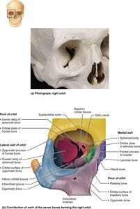
Special Features of the Orbits and Nasal Cavity
The Orbits
Bony cavities housing the eyes, cushioned by fatty tissue.
Formed by parts of seven bones: frontal, sphenoid, zygomatic, maxillary, palatine, lacrimal, and ethmoid.
Contain openings for nerves and blood vessels (superior/inferior orbital fissures, optic canal).
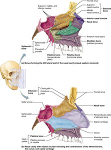
The Nasal Cavity
Constructed of bone and hyaline cartilage.
Roof: cribriform plates of ethmoid; lateral walls: conchae and perpendicular plates; floor: palatine processes of maxillae and palatine bones.
Divided by the nasal septum (vomer and perpendicular plate of ethmoid, plus septal cartilage).
Lined with mucosa to warm, moisten, and filter air; conchae increase turbulence for air filtration.
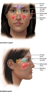
Paranasal Sinuses
Structure and Function
Located in the frontal, sphenoid, ethmoid, and maxillary bones.
Air-filled, mucosa-lined spaces that lighten the skull, warm and humidify air, and enhance voice resonance.
Self-Assessment and Clinical Connections
Key Questions
Which bones are the keystone bones of the facial skeleton? Maxillae.
What bone forms the bulk of the orbit floor? Maxilla. The sense organ found in the orbit is the eye.
What is the only freely movable joint in the skull? Temporomandibular joint (mandible).
What is the likely cause of death if the hyoid bone is fractured? Strangulation.
What process forms most skull bones in the embryo? Intramembranous ossification from mesenchymal connective tissue.
Clinical Note
Mastoiditis: Infection of the mastoid air cells, which can spread to the brain due to thin bony separation.
Summary Table: Major Cranial and Facial Bones and Markings
Bone | Key Markings | Main Functions |
|---|---|---|
Frontal | Supraorbital foramen, glabella, frontal sinus | Forehead, roof of orbits, anterior cranial fossa |
Parietal | Coronal, sagittal, lambdoid, squamous sutures | Superior/lateral skull |
Occipital | Foramen magnum, occipital condyles, nuchal lines | Posterior skull, cranial base |
Temporal | Zygomatic process, mastoid/styloid process, external acoustic meatus | Inferolateral skull, cranial base |
Sphenoid | Sella turcica, optic canal, pterygoid processes | Keystone of cranium |
Ethmoid | Cribriform plate, crista galli, perpendicular plate | Medial orbit, nasal cavity, septum |
Mandible | Body, ramus, alveolar process, mental foramen | Lower jaw, teeth anchor |
Maxilla | Alveolar/palatine processes, infraorbital foramen | Upper jaw, hard palate |
Zygomatic | Temporal/frontal/maxillary processes | Cheekbones, orbit margin |
Nasal | Articulates with frontal, maxilla, ethmoid | Bridge of nose |
Lacrimal | Lacrimal fossa | Medial orbit wall, tear drainage |
Palatine | Horizontal/perpendicular plates | Posterior hard palate, nasal cavity |
Vomer | Plow-shaped | Nasal septum |
Inferior Nasal Concha | Curved bone | Lateral nasal cavity |
