 Back
BackThe Somatic Nervous System: Sensory and Motor Pathways
Study Guide - Smart Notes
 Tailored notes based on your materials, expanded with key definitions, examples, and context.
The Somatic Nervous System
Introduction
The somatic nervous system is responsible for voluntary control of body movements via skeletal muscles and for processing sensory information from the external environment and internal body conditions. This system includes sensory receptors, sensory and motor pathways, and cortical processing centers.
Sensory Perception
Overview of Sensory Perception
Sensory receptors convert environmental or internal stimuli into electrochemical signals known as action potentials.
These action potentials are relayed to the central nervous system (CNS) for processing.
Perception is the conscious awareness of a sensation, while sensation is the activation of sensory receptor cells.
Not all sensations are perceived; many internal stimuli are processed subconsciously.
Example: Odorants from freshly baked cookies activate olfactory receptors, leading to the conscious perception of the smell.
Sensory Receptors
Types of Sensory Receptors
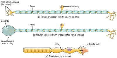
Sensory receptors are specialized to detect specific types of stimuli and can be classified based on their structure and function.
Free nerve endings: Dendrites embedded in tissues, responsible for detecting pain (nociceptors) and temperature (thermoreceptors).
Encapsulated nerve endings: Sensory neurons with endings surrounded by connective tissue, such as mechanoreceptors for pressure and touch.
Specialized receptor cells: Non-neuronal cells that detect specific stimuli, such as photoreceptors in the retina for light.
Chemoreceptors: Detect chemical stimuli, such as odorants (smell) and tastants (taste).


Sensory Pathways
Organization of Sensory Pathways
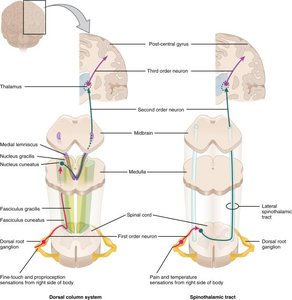
Sensory information from the body is transmitted to the CNS via spinal nerves, which split into dorsal and ventral roots near the spinal cord.
The dorsal root contains only sensory neuron axons; their cell bodies are in the dorsal root ganglion.
Sensory information travels to the brain via ascending tracts, typically involving three consecutive neurons (first-order, second-order, and third-order neurons).
Many sensory pathways are contralateral, meaning the right side of the body is processed by the left side of the brain and vice versa. The location of decussation (crossing over) varies by pathway.

Cortical Processing
Somatosensory Cortex and Homunculus
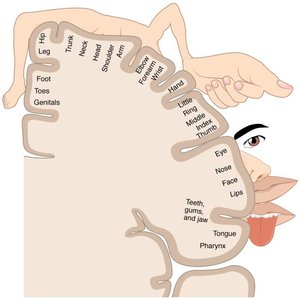
The primary somatosensory cortex is located in the postcentral gyrus of the parietal lobe. It processes touch, proprioception, nociception, and temperature information.
The sensory homunculus is a visual representation of the cortical area dedicated to processing sensory input from different body regions.
Body areas with higher sensitivity (e.g., hands, lips) occupy larger cortical regions.
Descending Pathways
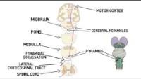
Motor Pathways and the Corticospinal Tract
Motor commands from the brain are transmitted to skeletal muscles via descending pathways.
The ventral roots of spinal nerves contain axons of motor neurons, whose cell bodies are in the ventral horns of the spinal cord.
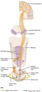
The corticospinal tract is the main descending pathway for voluntary motor control, involving two consecutive neurons (upper and lower motor neurons).
Most corticospinal tract axons are contralateral, crossing over at the pyramidal decussation in the medulla.
The lateral corticospinal tract controls appendicular muscles, while the anterior corticospinal tract controls trunk muscles. Some anterior tract axons are ipsilateral.
Motor commands end at the neuromuscular junction, causing muscle contraction.

Summary Table: Sensory and Motor Pathways
Pathway | Direction | Number of Neurons | Decussation | Function |
|---|---|---|---|---|
Ascending (Sensory) | Body to Brain | 3 | Varies by tract | Touch, pain, temperature, proprioception |
Descending (Motor) | Brain to Body | 2 | Medullary pyramids (mostly) | Voluntary muscle control |
Key Terms
Action potential: An electrical signal that travels along the membrane of a neuron.
Decussation: The crossing over of nerve fibers from one side of the CNS to the other.
Homunculus: A distorted representation of the human body, based on neurological "map" of the areas and proportions of the brain dedicated to processing motor or sensory functions.
Neuromuscular junction: The synapse between a motor neuron and a skeletal muscle fiber.
