 Back
The Special Senses: Smell, Taste, Vision, Hearing, and Equilibrium
Study Guide - Smart Notes
 Tailored notes based on your materials, expanded with key definitions, examples, and context.
The Special Senses
Overview of the Special Senses
The special senses include smell, taste, vision, hearing, and equilibrium. These senses are housed in complex sensory organs and are essential for perceiving the environment and maintaining homeostasis.
Smell (Olfaction) and Taste (Gustation) are chemical senses, relying on chemoreceptor cells.
Vision is processed by the eyes, which contain more than half of the body's sensory receptors.
Hearing and Equilibrium are mediated by the ear, which contains specialized structures for sound and balance.
Chemical Senses: Olfaction and Gustation
Olfactory Epithelium and Olfactory Pathway
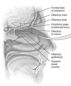
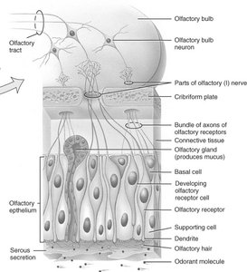
Olfaction is the sense of smell, detected by olfactory receptors located in the superior portion of the nasal cavity. These receptors are bipolar neurons with cilia (olfactory hairs) that combine to form the olfactory (I) nerve. Basal cells act as stem cells, replacing olfactory receptors monthly, while olfactory glands produce mucus.
Odorant molecules bind to receptor proteins, triggering nerve impulses.
Olfactory signals project to the cerebral cortex and limbic system, evoking strong emotional reactions.
Odor Thresholds and Adaptation
Low threshold: Only a few molecules are needed to trigger a response (e.g., methyl mercaptan in natural gas).
Adaptation: Sensitivity decreases rapidly—50% in 1 second, complete in 1 minute for strong odors.
Gustatory Sensation: Taste
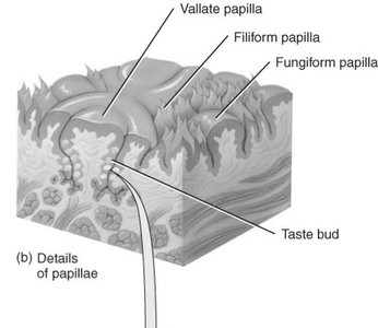
Taste is a chemical sense requiring molecules to be dissolved for detection. There are five primary taste sensations: sour, sweet, bitter, salty, and umami (savory/meaty/glutamate). Other tastes are combinations of these primary sensations plus olfaction. Taste buds are found on the sides of papillae on the tongue. 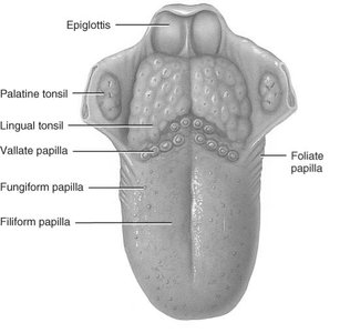
Anatomy and Physiology of Taste Buds
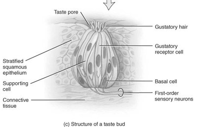
Taste buds are oval bodies consisting of about 50 receptor cells, basal cells, and supporting cells. Each receptor cell contains a single gustatory hair (microvillus) that extends through a taste pore.
Mechanism: Dissolved substances contact gustatory hairs, leading to receptor potentials, neurotransmitter release, and nerve impulse formation.
Thresholds: Most sensitive to bitter (often poisons), least sensitive to salty and sweet.
Vision
Accessory Structures of the Eye
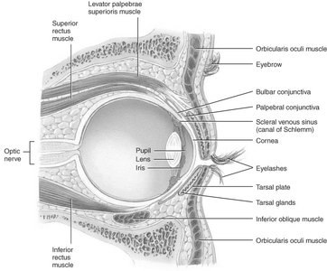
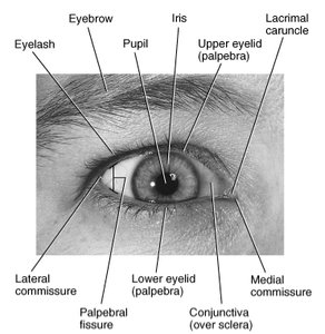
The eye is protected and maintained by several accessory structures, including eyelids, conjunctiva, and extrinsic eye muscles.
Eyelids, Eyelashes, and Eyebrows: Protect from foreign objects, perspiration, and sunlight.
Tarsal glands: Sebaceous glands that prevent eyelids from sticking and slow tear evaporation.
Lacrimal Apparatus
The lacrimal apparatus produces and distributes tears, which contain lysozyme, a bactericidal enzyme. Tears are spread over the eye by blinking and drain into the nasal cavity. 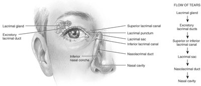
Three Tunics (Layers) of the Eyeball
The eyeball consists of three main layers:
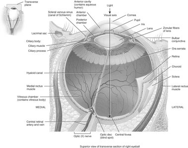
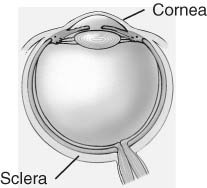
Fibrous Tunic (outer layer): Includes the cornea (transparent, focuses light) and sclera (white of the eye).
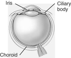
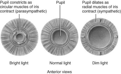
Vascular Tunic (middle layer): Includes the choroid (nutrients and melanocytes), ciliary body (muscle and process), and iris (colored portion, regulates light).
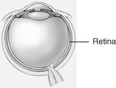
Nervous Tunic (inner layer): The retina, containing photoreceptors and neurons.
Lens
The lens is avascular and composed of crystallin proteins arranged in layers. It focuses light on the retina and is connected to the ciliary body. With aging, the lens loses elasticity, resulting in presbyopia. 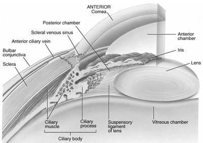
Nervous Tunic: Retina
The retina lines the posterior 3/4 of the eyeball and contains the optic disc (blind spot), blood vessels, and macula lutea (central fovea). The retina is susceptible to diseases such as hypertension, macular disease, diabetes, and trauma (detached retina). 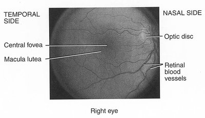
Layers of the Retina
The retina consists of a pigmented epithelium (absorbs stray light) and three layers of neurons: photoreceptor layer, bipolar neuron layer, and ganglion neuron layer. 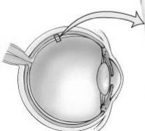
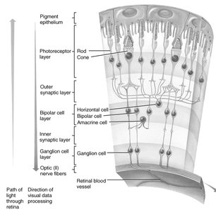
Photoreceptors: Rods and Cones
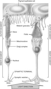
Rods: Rod-shaped, detect shades of gray in dim light, 120 million cells, distributed along periphery.
Cones: Cone-shaped, sharp color vision in bright light, 6 million cells, concentrated in central fovea.
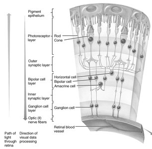
Pathway of Nerve Signal in Retina
Light penetrates the retina, where rods and cones transduce light into action potentials. These excite bipolar cells, which in turn excite ganglion cells. Axons of ganglion cells form the optic (II) nerve, which transmits signals to the thalamus and primary visual cortex. 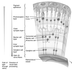
Cavities of the Interior of Eyeball
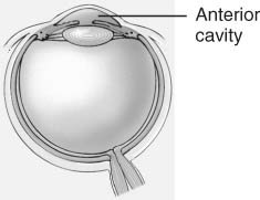
Anterior cavity: Filled with aqueous humor, produced by ciliary body and drained into canal of Schlemm.
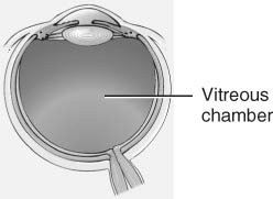
Vitreous chamber: Filled with vitreous body (jellylike), formed during embryonic life.
Intraocular Pressure
Intraocular pressure is controlled mainly by aqueous humor and maintains the shape of the eyeball. Increased pressure (glaucoma) can cause degeneration of the retina and blindness.
Major Processes of Image Formation
Constriction/dilation of the pupil: Controls the amount of light entering the eye.
Refraction of light: By cornea and lens.
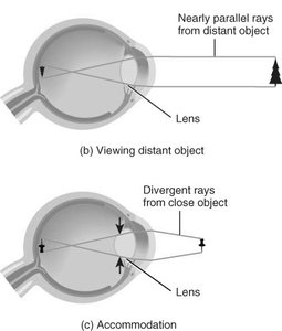
Accommodation of the lens: Changing shape of lens to focus light.
Refraction and Accommodation
Images focused on the retina are inverted and reversed.
Accommodation increases the curvature of the lens for near objects.
Correction for Refraction Problems
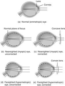
Emmetropia: Normal vision.
Myopia (nearsighted): Eyeball too long; corrected with concave lenses.
Hypermetropia (farsighted): Eyeball too short; corrected with convex lenses.
Astigmatism: Corneal surface wavy; parts of image out of focus.
Physiology of Vision
The first step in vision transduction is the absorption of light by photopigments in rods and cones. Photopigments undergo structural changes upon light absorption.
Retinal: Light-absorbing part of photopigments.
Opsin: Protein component; four types exist.
Rhodopsin: Photopigment in rods.
Cones: Three types, each absorbing different wavelengths (colors).
Regeneration of Bleached Photopigments
Bleaching and regeneration of photopigments account for light and dark adaptation.
Rhodopsin regeneration takes 30–40 minutes; cones regenerate in 90 seconds.
Rods contribute little to daylight vision due to rapid bleaching.
Color Blindness and Night Blindness
Color blindness: Inability to distinguish certain colors, usually due to inherited deficiency in cone photopigments.
Night blindness (nyctalopia): Difficulty seeing in low light, often due to vitamin A deficiency.
Hearing and Equilibrium
Anatomy of the Ear Region
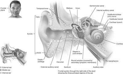
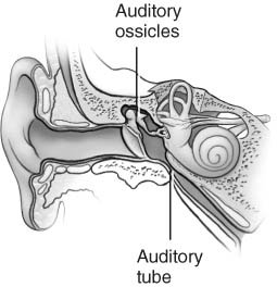
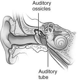
The ear is divided into three regions: external, middle, and internal ear.
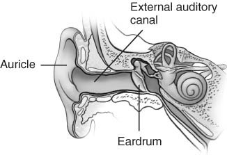
External ear: Collects sound waves (auricle, external auditory canal, tympanic membrane).
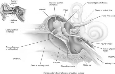
Middle ear: Air-filled cavity with three ossicles (malleus, incus, stapes) and auditory (Eustachian) tube.
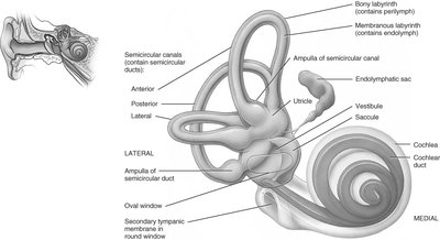
Inner ear: Fluid-filled labyrinth containing cochlea (hearing) and vestibule/semicircular ducts (balance).
Physiology of Hearing
Sound waves are collected by the auricle, vibrate the eardrum, and are transmitted by ossicles to the oval window.
Fluid pressure waves in the cochlea move hair cells against the tectorial membrane, bending microvilli and producing receptor potentials.
Cochlear Anatomy
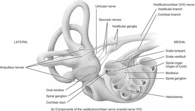
The cochlea contains three fluid-filled channels: scala vestibuli, scala tympani, and cochlear duct. Vibrations from the stapes are transmitted into the fluid of the scala vestibuli, affecting hair cells in the cochlear duct.
Organ of Corti
The organ of Corti is the primary sense organ for hearing. Pressure fluctuations move hair cells against the tectorial membrane, generating nerve impulses in the cochlear nerve.
Sound Waves, Pitch, and Volume
Frequency: Perceived as pitch; higher frequency = higher pitch.
Amplitude: Perceived as volume; greater amplitude = louder sound.
Deafness
Nerve deafness: Damage to hair cells or cranial nerve VIII.
Conduction deafness: Vibrations not conducted to hair cells (e.g., perforated eardrum, otosclerosis).
Cochlear Implants
Cochlear implants can restore hearing by translating sounds into electrical stimulation of the vestibulocochlear nerve.
Equilibrium (Balance)
Vestibular Apparatus
The vestibular apparatus includes semicircular ducts with ampulla and the vestibule (utricle and saccule).
Physiology of Equilibrium
Static equilibrium: Maintains head position relative to gravity (macula receptors in saccule and utricle).
Dynamic equilibrium: Maintains head position during movement (crista receptors in ampulla of semicircular ducts).
The Macula
The maculae of the utricle and saccule are sense organs for static equilibrium. Hair cells with stereocilia are embedded in a gelatinous otolithic membrane containing calcium carbonate crystals (otoliths) that move with head position.
Detection of Position and Rotational Movement
Movement of stereocilia releases neurotransmitter onto vestibular branches of the vestibulocochlear nerve (VIII). The cristae in the ampulla of semicircular ducts detect rotational movement, with hair cells covered by a gelatinous cupula.
Aging and Disorders of the Special Senses
Aging
Loss of olfactory and gustatory receptors after age 50.
Presbycusis (hearing loss), tinnitus, presbyopia, cataracts, macular disease, detached retina, glaucoma, decreased tear production, reduced vision sharpness, depth, and color perception.
Disorders: Homeostatic Imbalances
Otitis media: Acute infection of the middle ear, primarily by bacteria. Characterized by pain, malaise, fever, and bulging of the eardrum. More common in children.
Special Sense | Receptor Type | Location | Main Function |
|---|---|---|---|
Smell (Olfaction) | Olfactory receptors (bipolar neurons) | Superior nasal cavity | Detects odorants |
Taste (Gustation) | Gustatory receptor cells | Taste buds on papillae | Detects dissolved chemicals |
Vision | Rods and cones | Retina | Detects light, color, and movement |
Hearing | Hair cells | Cochlea | Detects sound vibrations |
Equilibrium | Hair cells | Vestibule and semicircular ducts | Detects head position and movement |
Additional info: Academic context was added to clarify mechanisms, anatomical relationships, and clinical relevance for each special sense.
