 Back
BackThe Special Senses: Structure and Function in Human Anatomy & Physiology
Study Guide - Smart Notes
 Tailored notes based on your materials, expanded with key definitions, examples, and context.
Overview of the Special Senses
Introduction to Special Senses
The special senses are specialized sensory modalities that detect specific stimuli through dedicated organs located primarily in the head. These senses include smell (olfaction), taste (gustation), vision, hearing (audition), and vestibular sensation (balance). Each sense is associated with unique receptor cells and neural pathways that transmit information to the central nervous system for processing.
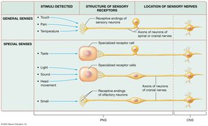
Comparison of General and Special Senses
General senses and special senses differ in the types of stimuli they detect, the structure of their sensory receptors, and the location of their sensory nerves.
Aspect | General Senses | Special Senses |
|---|---|---|
Stimuli Detected | Touch, pain, temperature | Light, sound, head movement, chemicals (taste/smell) |
Receptor Structure | Receptive ends of sensory neurons | Specialized receptor cells (except olfaction) |
Location of Sensory Nerves | Axons in spinal and cranial nerves | Axons in cranial nerves (all in the head) |
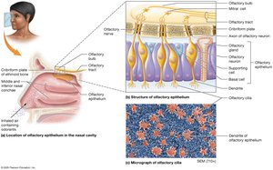
Olfaction (Smell)
Structures of Olfaction
Olfaction enables the detection of odorants (chemicals in the air). Humans can detect up to 400,000 odors, with most being unpleasant. The olfactory epithelium is located in the superior region of each nasal cavity and contains three main cell types:
Olfactory Neurons: Modified neurons with chemoreceptors that detect odorants. Their axons form the olfactory nerve, which transmits signals to the olfactory bulb and tract.
Basal Cells: Stem cells that replace olfactory neurons every 30–60 days.
Supporting Cells: Columnar cells that provide structural support.
Clinical Connection: Anosmia and COVID-19
Anosmia is the loss of the sense of smell and is an early symptom of COVID-19. It is likely due to viral effects on olfactory neuron nuclei, reducing receptor production. Most cases are temporary, with recovery in weeks to years.
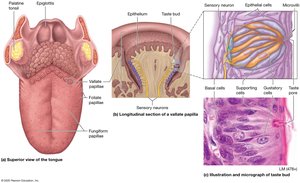
Gustation (Taste)
Structures and Types of Papillae
Gustation involves the detection of chemicals by taste buds, which are clusters of receptor and supporting cells on the tongue and oral cavity. The tongue is covered with papillae of various types:
Vallate (Circumvallate) Papillae: Large, dome-shaped, with many taste buds.
Fungiform Papillae: Mushroom-shaped, with a few taste buds each.
Foliate Papillae: Ridges on the sides of the tongue, taste buds present mainly in childhood.
Filiform Papillae: Long, thin, lack taste buds but detect texture and temperature.
Taste Buds and Cell Types
Taste buds are found on the lateral surfaces of papillae and contain:
Gustatory (Taste) Cells: Specialized epithelial cells with microvilli that synapse with sensory neurons (via facial, glossopharyngeal, or vagus nerves). Lifespan: 10–14 days.
Basal Cells: Stem cells for gustatory cell replacement.
Supporting Cells: Provide structural support.
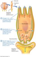
Physiology of Gustation
Taste sensations are based on the detection of chemicals and the combination of activated receptors. The five primary taste sensations are:
Sweet: Simple sugars (e.g., glucose, fructose)
Sour: Hydrogen ions (e.g., citric acid)
Salty: Metal ions (e.g., sodium, potassium)
Bitter: Alkaloids (e.g., in coffee, rancid foods)
Umami: Glutamate or other amino acids (savory)
Vision
Accessory Structures of the Eye
The eye is protected and supported by several accessory structures:
Eyelids (Palpebrae): Protect the eye and distribute tears.
Eyebrows: Prevent sweat from entering the eyes.
Eyelashes: Trigger the blink reflex.
Conjunctiva: Thin membrane covering the anterior eye; inflammation is called conjunctivitis.
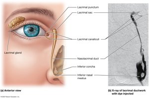
Lacrimal Apparatus: Produces and drains tears via the lacrimal gland, sac, and nasolacrimal duct.
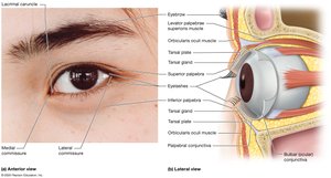
Structure of the Eyeball
The eyeball is a hollow sphere with three main tissue layers (tunics):
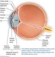
Fibrous Layer: Outermost; includes the sclera (white) and cornea (transparent, allows light entry).
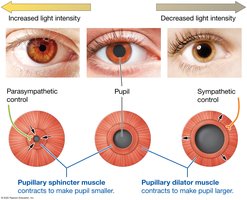
Vascular Layer: Middle; includes the choroid (blood supply), ciliary body (smooth muscle, suspensory ligaments), and iris (pigmented, controls pupil size).
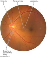
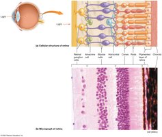
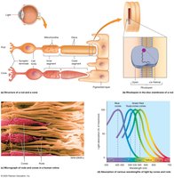
Neural Layer (Retina): Innermost; contains photoreceptors (rods and cones), fovea centralis (high acuity), and optic disc (blind spot).
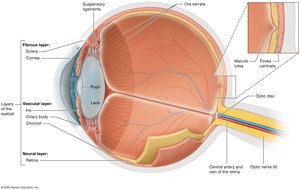
Chambers and Lens of the Eye
The lens focuses light on the retina. The posterior cavity contains vitreous humor (gel-like), while the anterior cavity contains aqueous humor (fluid). These maintain eye shape and optical properties.
Common Eye Disorders
Cataracts: Clouding of the lens, leading to blindness; treated by lens replacement.
Glaucoma: Increased intraocular pressure damages the retina and optic nerve; managed with medication or surgery.
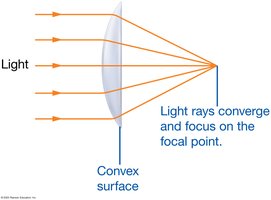
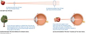
Principles of Light and Refraction
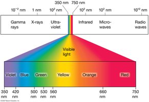
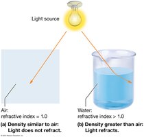
Vision is based on the perception of electromagnetic radiation (visible light, 400–700 nm). Light rays bend (refract) when passing through different media, focusing on the retina. The cornea provides most refraction; the lens fine-tunes focus, especially for near objects (accommodation).
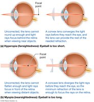
Errors of Refraction
Presbyopia: Age-related loss of accommodation; corrected with reading glasses.
Hyperopia (Farsightedness): Eyeball too short or cornea too flat; corrected with convex lenses.
Myopia (Nearsightedness): Eyeball too long or cornea too curved; corrected with concave lenses.
Astigmatism: Irregular curvature of lens/cornea; corrected with special lenses or LASIK surgery.
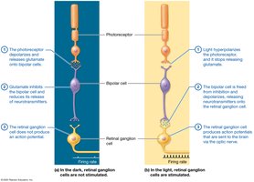
Photoreceptors and the Retina
The retina contains two main types of photoreceptors:
Rods: Sensitive to low light, black-and-white vision, peripheral vision; contain rhodopsin.
Cones: Responsible for color and high-acuity vision; concentrated in the fovea; contain iodopsin (three types for red, green, blue).
Color Blindness
Color blindness is a genetic disorder affecting cone pigments, most commonly red-green. It is more prevalent in males due to its sex-linked inheritance.
Image Processing by the Retina
Most retinal ganglion cells receive input from multiple photoreceptors, except in the fovea, where one-to-one connections allow for sharp vision. Peripheral vision is less detailed due to convergence.
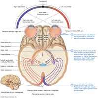
The Visual Pathway
Visual information from each eye is processed in the brain to create depth perception (stereoscopic vision). The visual field is the area seen by each eye, with overlap allowing for binocular vision.
Hearing and Equilibrium
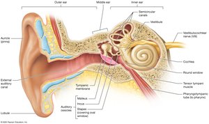
Anatomy of the Ear
The ear is divided into three regions:
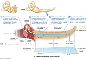
Outer Ear: Auricle (pinna), external auditory canal, tympanic membrane; funnels and transmits sound.
Middle Ear: Air-filled cavity with auditory ossicles (malleus, incus, stapes) and pharyngotympanic tube; transmits and amplifies sound.
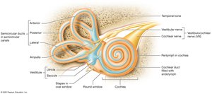
Inner Ear: Bony and membranous labyrinths; contains cochlea (hearing) and vestibular apparatus (balance).
Otitis Media
Otitis media is inflammation of the middle ear, common in children due to shorter, more horizontal auditory tubes. It can cause pain, fever, and hearing loss, and may require antibiotics.
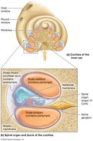
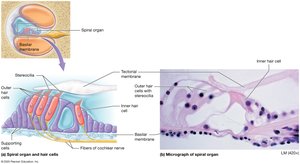
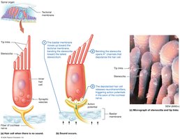
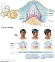
Inner Ear: Cochlea and Vestibular System
The cochlea is responsible for hearing and contains the spiral organ (Organ of Corti), which houses hair cells that transduce sound into neural signals. The vestibule and semicircular canals detect head position and movement for balance.
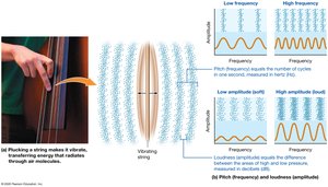
Principles of Sound
Sound waves are vibrations of air molecules. Pitch (frequency) is measured in hertz (Hz), and loudness (amplitude) in decibels (dB). Humans detect sounds from 20 Hz to 20,000 Hz.
Vestibular Sensation (Equilibrium)
Static and Dynamic Equilibrium
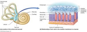
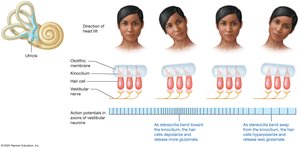
Equilibrium depends on input from vision, proprioceptors, and the vestibular system. Static equilibrium is monitored by the utricle and saccule, which detect head position and linear acceleration via hair cells embedded in the otolithic membrane. Dynamic equilibrium involves the semicircular ducts, which detect rotational movements.
Motion Sickness
Motion sickness results from conflicting sensory information between the eyes and vestibular system. Symptoms include nausea and dizziness, and can be alleviated with medication or behavioral strategies.
Summary Table: Special Senses and Their Receptors
Sense | Receptor Type | Location | Main Function |
|---|---|---|---|
Olfaction | Chemoreceptors (neurons) | Olfactory epithelium (nasal cavity) | Detects odorants |
Gustation | Chemoreceptors (epithelial cells) | Taste buds (tongue, oral cavity) | Detects tastants |
Vision | Photoreceptors (rods/cones) | Retina (eye) | Detects light |
Hearing | Mechanoreceptors (hair cells) | Spiral organ (cochlea) | Detects sound waves |
Equilibrium | Mechanoreceptors (hair cells) | Vestibule, semicircular canals (inner ear) | Detects head position/movement |
