 Back
BackThe Special Senses: Structure and Function
Study Guide - Smart Notes
 Tailored notes based on your materials, expanded with key definitions, examples, and context.
Chapter 15: The Special Senses
Overview of the Special Senses
The special senses include olfaction (smell), gustation (taste), vision, audition (hearing), and equilibrium (balance). These senses rely on specialized sensory organs and distinct neural pathways to convey specific types of information to the brain, distinguishing them from the general senses such as touch, pain, and temperature.
Special senses: Olfaction, gustation, vision, audition, equilibrium
General senses: Touch, pain, temperature, proprioception
Key brain areas: Each special sense is processed in specific regions of the brain, such as the olfactory cortex, gustatory cortex, visual cortex, auditory cortex, and vestibular nuclei.
Sensory transduction is the process by which physical stimuli are converted into neural signals. This involves receptor cells detecting a stimulus and generating action potentials that travel to the brain for interpretation.
Module 15.2: Olfaction (Smell)
Olfactory Receptors and Pathways
Olfaction is the sense of smell, mediated by chemoreceptors located in the olfactory epithelium of the nasal cavity. These receptors detect airborne chemicals called odorants and transduce them into neural signals.
Olfactory epithelium: Contains olfactory receptor cells, supporting cells, and basal cells.
Olfactory bulb: The first relay station in the brain for olfactory information.
Pathway: Odorants bind to receptors, triggering action potentials that travel via the olfactory nerve to the olfactory bulb, then to the olfactory cortex and limbic system.
Importance: The connection to the limbic system explains why smells can evoke strong emotions and memories.
Module 15.3: Gustation (Taste)
Taste Buds and Gustatory Pathways
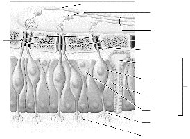
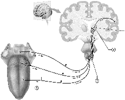
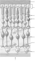
Gustation is the sense of taste, detected by taste buds located primarily on the tongue. Each taste bud contains gustatory (taste) cells that respond to chemicals dissolved in saliva.
Types of papillae: Vallate, fungiform, and foliate papillae house taste buds.
Primary tastes: Sweet, sour, salty, bitter, umami
Pathway: Gustatory cells generate action potentials that travel via cranial nerves VII, IX, and X to the medulla, thalamus, and finally the primary gustatory cortex.
Module 15.4: Anatomy of the Eye
Accessory Structures and Layers of the Eye
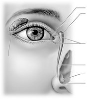
The eye is a complex organ with several accessory structures and three main layers. Accessory structures include the palpebrae (eyelids), conjunctiva, lacrimal gland, and nasolacrimal duct, which protect and lubricate the eye.
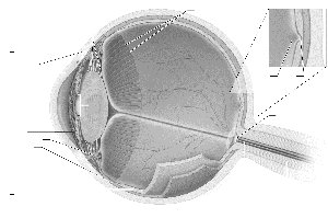
Fibrous layer: Sclera and cornea
Vascular layer: Choroid, ciliary body, iris
Neural layer: Retina, containing photoreceptors (rods and cones)
Main Features of the Retina
The retina contains several layers of cells, including photoreceptors, bipolar cells, and ganglion cells. The macula lutea and fovea centralis are specialized for high-acuity vision, while the optic disc is the site where the optic nerve exits the eye.
Module 15.5: Physiology of Vision
Light Transduction and Visual Pathways
Light entering the eye is focused by the cornea and lens onto the retina. Photoreceptors (rods and cones) convert light into electrical signals. Rods are sensitive to low light, while cones detect color and detail.
Accommodation: The lens changes shape to focus on near or distant objects.
Visual pathway: Signals travel from the retina through the optic nerve, cross at the optic chiasma, and reach the visual cortex.
Light/dark adaptation: The retina adjusts sensitivity to different lighting conditions.
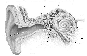
Module 15.6: Anatomy of the Ear
Regions and Structures of the Ear
The ear is divided into three main regions: outer, middle, and inner ear. Each region contains specialized structures for hearing and balance.
Outer ear: Auricle, external auditory canal
Middle ear: Tympanic membrane, auditory ossicles, pharyngotympanic tube
Inner ear: Bony labyrinth (cochlea, vestibule, semicircular canals), filled with perilymph and endolymph
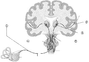
Module 15.7: Physiology of Hearing
Sound Transduction and Auditory Pathways
Sound waves are collected by the auricle, transmitted through the auditory canal, and vibrate the tympanic membrane. These vibrations are amplified by the ossicles and transmitted to the cochlea, where hair cells in the spiral organ convert mechanical energy into neural signals.
Pitch and loudness: Determined by the location and frequency of hair cell activation.
Auditory pathway: Signals travel via the vestibulocochlear nerve to the brainstem, thalamus, and auditory cortex.
Module 15.8: Vestibular Sensation (Equilibrium)
Static and Dynamic Equilibrium
The vestibular system detects head position and movement to maintain balance. Static equilibrium is sensed by the maculae of the utricle and saccule, while dynamic equilibrium is detected by the crista ampullaris in the semicircular canals.
Macula: Contains hair cells embedded in the otolithic membrane, which shifts with head movement.
Crista ampullaris: Contains hair cells in the ampulla of semicircular canals, detecting rotational movement.
Module 15.9: Integration of the Special Senses
Pathways and Integration in the Brain
Signals from the special senses are integrated in the frontal lobe and limbic system, allowing for a comprehensive perception of the environment. This integration is essential for behaviors such as memory formation, emotional responses, and coordinated movement.
Summary Table: Comparison of Special Senses
Sense | Receptor Type | Location | Main Brain Area |
|---|---|---|---|
Olfaction | Chemoreceptor | Olfactory epithelium | Olfactory cortex, limbic system |
Gustation | Chemoreceptor | Taste buds (tongue) | Gustatory cortex |
Vision | Photoreceptor | Retina | Visual cortex |
Audition | Mechanoreceptor | Cochlea | Auditory cortex |
Equilibrium | Mechanoreceptor | Vestibular apparatus | Vestibular nuclei, cerebellum |
Key Equations and Concepts
Refraction of light: (Snell's Law, where n is the refractive index)
Accommodation: (Lens equation, where f is focal length, d_o is object distance, d_i is image distance)
Additional info: Academic context and explanations have been expanded for clarity and completeness. Images included only where directly relevant to the described structures or pathways.
