 Back
The Special Senses: Structure and Function of the Eye
Study Guide - Smart Notes
 Tailored notes based on your materials, expanded with key definitions, examples, and context.
Introduction to the Special Senses
The special senses—olfaction, gustation, vision, equilibrium, and hearing—provide critical information about the environment. Vision, in particular, is the most relied-upon sense in humans, involving complex anatomical structures and intricate neural pathways. This chapter focuses on the anatomy and physiology of the eye, the organ responsible for vision, and the mechanisms underlying visual perception.
Accessory Structures of the Eye
Eyelids and Superficial Epithelium
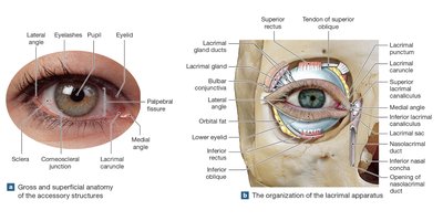
The accessory structures of the eye protect, lubricate, and support the eyeball. The eyelids (palpebrae) are extensions of the skin that blink to keep the eye moist and free from debris. The palpebral fissure is the gap between the upper and lower eyelids, while the medial and lateral canthi are the points where the eyelids meet. Eyelashes prevent foreign matter from reaching the eye, and tarsal glands secrete lipids to prevent eyelid adhesion. The conjunctiva is a mucous membrane covering the inner eyelids (palpebral conjunctiva) and the anterior surface of the eye (bulbar conjunctiva), extending to the cornea.
Conjunctivitis (pinkeye): Inflammation of the conjunctiva, often resulting in redness due to dilated blood vessels.
Lacrimal caruncle: Contains glands producing secretions that may accumulate during sleep.
Clinical terms: Chalazion (cyst from tarsal gland infection), sty (painful swelling from sebaceous gland infection).
The Lacrimal Apparatus
The lacrimal apparatus produces, distributes, and removes tears, which lubricate the eye, reduce friction, remove debris, and contain antibacterial enzymes (lysozyme). It consists of the lacrimal gland, lacrimal canaliculi, lacrimal sac, and nasolacrimal duct. Tears drain into the nasal cavity, explaining why crying leads to a runny nose.
Lacrimal gland: Produces most of the tear volume; located superior and lateral to the eyeball.
Lacrimal puncta: Small pores that drain tears into the canaliculi.
Nasolacrimal duct: Delivers tears to the nasal cavity.
Anatomy of the Eyeball
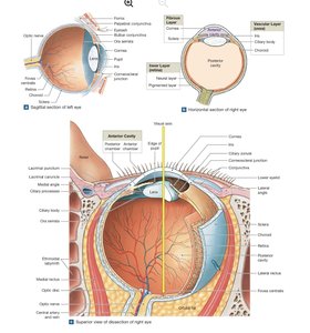
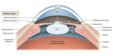
The eyeball is a nearly spherical organ with three main layers: the fibrous layer, vascular layer (uvea), and inner layer (retina). It contains two cavities: the anterior cavity (filled with aqueous humor) and the posterior cavity (filled with vitreous body).
The Fibrous Layer
Sclera: The white, tough outer layer providing protection and muscle attachment.
Cornea: Transparent anterior portion allowing light entry; avascular and highly sensitive.
Corneal limbus: Junction between sclera and cornea.
Clinical note: Corneal injuries can cause blindness; corneal transplants are common due to the lack of blood vessels.
The Vascular Layer (Uvea)
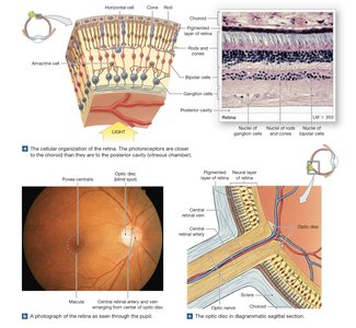
Iris: Pigmented structure controlling pupil diameter via the dilator and sphincter pupillae muscles. Eye color is determined by melanocyte density and pigment distribution.
Ciliary body: Contains the ciliary muscle and processes; attaches to the lens via the ciliary zonule (suspensory ligament) and controls lens shape.
Choroid: Vascular layer supplying nutrients and oxygen to the retina; contains melanocytes to absorb stray light.
The Inner Layer: The Retina
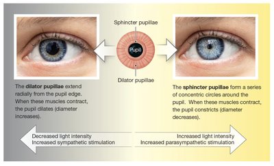
The retina consists of a pigmented layer (absorbs stray light) and a neural layer (contains photoreceptors and neurons). The macula is the region of highest cone density, with the fovea centralis at its center for sharpest vision. The optic disc is the origin of the optic nerve and forms the blind spot due to the absence of photoreceptors.
Photoreceptors: Rods (dim light, no color) and cones (color, high acuity).
Bipolar cells: Relay signals from photoreceptors to ganglion cells.
Ganglion cells: Axons form the optic nerve.
Horizontal and amacrine cells: Modulate signal transmission and sensitivity.
Chambers and Fluids of the Eye
Aqueous Humor
Aqueous humor is produced by the ciliary processes, circulates through the anterior cavity, and drains via the scleral venous sinus. It provides nutrients, removes wastes, and maintains intraocular pressure (normally 12–21 mm Hg).
Clinical note: Blocked drainage leads to glaucoma, increasing pressure and risking optic nerve damage.
Vitreous Body
The vitreous body is a gelatinous mass in the posterior cavity, stabilizing the eye's shape and supporting the retina. It is not replaced after development.
The Lens
The lens is a transparent, biconvex structure that focuses light onto the retina by changing shape (accommodation). It is composed of lens fibers filled with crystallins. Loss of transparency leads to cataracts, which may require surgical replacement.
Photoreceptors and Visual Pigments
Structure of Rods and Cones
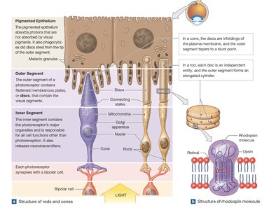
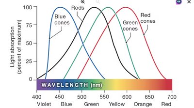
Rods and cones contain visual pigments in their outer segments. Rods contain rhodopsin, while cones contain photopsins sensitive to different wavelengths (blue, green, red).
Rods: High sensitivity, no color discrimination, function in low light.
Cones: Color vision, high acuity, require bright light.
Light Absorption and Color Vision
Each type of cone is most sensitive to a specific wavelength range. The combination of cone responses allows for color discrimination. Rods are most sensitive to wavelengths around 500 nm (green-blue).
Phototransduction: Bleaching and Regeneration
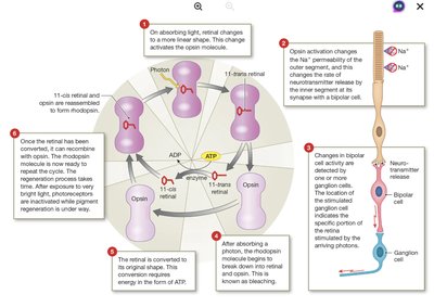
When a photon is absorbed, retinal changes shape, activating opsin and initiating a cascade that alters membrane permeability and neurotransmitter release. Rhodopsin is then bleached and must be regenerated, a process requiring ATP.
Bleaching: Separation of retinal and opsin after light absorption.
Regeneration: Enzymatic conversion of retinal back to its original form and recombination with opsin.
Adaptation and Night Blindness
Dark adaptation occurs when visual pigments are fully regenerated, increasing sensitivity. Light adaptation involves bleaching, reducing sensitivity. Vitamin A deficiency impairs pigment regeneration, leading to night blindness (nyctalopia).
Color Vision and Color Blindness
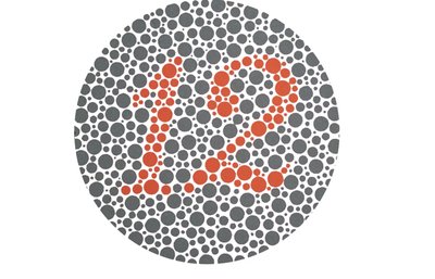
Color vision depends on the integration of signals from blue, green, and red cones. Color blindness results from the absence or malfunction of one or more cone types, most commonly red-green color blindness. Standard tests use colored dot patterns to assess color discrimination.
Visual Pathways and Processing
Retinal Processing
Photoreceptors synapse with bipolar cells, which in turn synapse with ganglion cells. Rods exhibit high convergence, leading to less precise vision, while cones have little convergence, allowing for high acuity. Ganglion cells are classified as M cells (motion, general form) and P cells (fine detail, color).
Central Visual Pathways
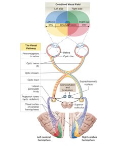
Axons of ganglion cells form the optic nerve, which partially decussates at the optic chiasm. Visual information is relayed to the lateral geniculate body of the thalamus and then to the visual cortex. Some fibers project to the superior colliculi and hypothalamus for reflex and circadian rhythm regulation.
Field of vision: Overlap of visual fields from both eyes enables depth perception.
Visual cortex: Processes and integrates visual information, creating a spatial map of the visual field.
Summary Table: Layers and Functions of the Eye
Layer | Main Structures | Functions |
|---|---|---|
Fibrous Layer | Sclera, Cornea | Protection, support, muscle attachment, light entry |
Vascular Layer (Uvea) | Iris, Ciliary body, Choroid | Blood supply, light regulation, lens shape, aqueous humor production |
Inner Layer (Retina) | Pigmented layer, Neural layer (rods, cones, bipolar cells, ganglion cells) | Light absorption, phototransduction, signal processing |
Key Equations
Focusing by the lens (Lens equation):
Where f is the focal length, d_o is the object distance, and d_i is the image distance.
Intraocular pressure (IOP): Normal range: 12–21 mm Hg
Clinical Correlations
Glaucoma: Increased intraocular pressure damages the optic nerve, leading to vision loss.
Cataracts: Clouding of the lens, impairing vision; treated by surgical replacement.
Diabetic retinopathy: Retinal blood vessel damage due to diabetes, leading to vision loss.
Detached retina: Separation of neural and pigmented layers, requiring prompt surgical intervention.
