 Back
BackThe Special Senses: Structure, Function, and Disorders
Study Guide - Smart Notes
 Tailored notes based on your materials, expanded with key definitions, examples, and context.
Chapter 15: The Special Senses
Introduction to Special Senses
The special senses include sight, hearing, equilibrium, smell, and taste. Each sense gathers unique sensory information, which is processed in specialized areas of the cerebrum and influences motor output. These senses are essential for interacting with the environment and maintaining homeostasis.
15-1 The Eye and Vision
Accessory Structures of the Eye
Eyelids: Protect the eye and help circulate fluids by blinking.
Eyelashes: Trap debris and prevent large particles from entering the eye.
Tarsal glands: Modified sebaceous glands that produce an oily secretion to lubricate the eye.
Ciliary glands: Modified sweat glands located between the eyelashes.
Conjunctiva: Membrane lining the eyelids and connecting to the outer surface of the eye; secretes mucus to keep the eye moist.
Lacrimal apparatus: Produces and drains tears, protecting, moistening, and lubricating the eye. Tears contain a dilute salt solution, mucus, antibodies, and lysozyme (an enzyme that destroys bacteria).
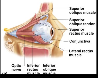
Extrinsic eye muscles: Six muscles that attach to the outer surface of the eye and produce eye movements.
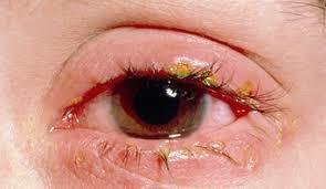
Conjunctivitis (Pinkeye)
A highly contagious infection of the conjunctiva, resulting in redness and discharge.
Lacrimal Apparatus Anatomy
Lacrimal gland: Produces lacrimal fluid (tears).
Excretory ducts: Carry fluid from the gland to the eye surface.
Lacrimal canaliculi: Drain fluid medially from the eye.
Lacrimal sac: Passageway for fluid toward the nasal cavity.
Nasolacrimal duct: Empties fluid into the nasal cavity.
Extrinsic Eye Muscles
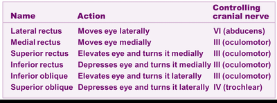
Six muscles control eye movement: lateral rectus, medial rectus, superior rectus, inferior rectus, inferior oblique, and superior oblique.
Each muscle is innervated by specific cranial nerves.
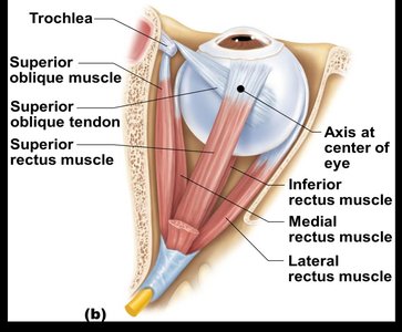
Name | Action | Controlling Cranial Nerve |
|---|---|---|
Lateral rectus | Moves eye laterally | VI (abducens) |
Medial rectus | Moves eye medially | III (oculomotor) |
Superior rectus | Elevates eye and turns it medially | III (oculomotor) |
Inferior rectus | Depresses eye and turns it medially | III (oculomotor) |
Inferior oblique | Elevates eye and turns it laterally | III (oculomotor) |
Superior oblique | Depresses eye and turns it laterally | IV (trochlear) |
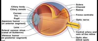
15-2 Structure of the Eye
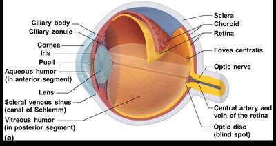
Fibrous Layer
Sclera: White, connective tissue layer; the "white of the eye."
Cornea: Transparent, anterior portion of the sclera; allows light to pass through and repairs itself easily.
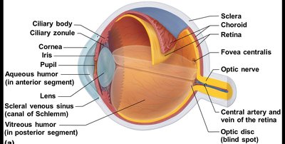
Vascular Layer
Choroid: Blood-rich nutritive layer; contains dark pigments to prevent light scattering.
Ciliary body: Smooth muscle attached to the lens by the ciliary zonule (suspensory ligaments).
Iris: Pigmented muscle that regulates the amount of light entering the eye; contains the pupil (opening).
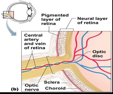
Sensory Layer (Retina)
Outer pigmented layer: Absorbs light and prevents scattering.
Inner neural layer: Contains photoreceptors (rods and cones).
Fovea centralis: Area of greatest visual acuity, contains only cones.
Optic disc (blind spot): Where the optic nerve exits; no photoreceptors present.

15-3 Photoreceptors: Rods and Cones
Rods
Located toward the edges of the retina.
Enable vision in dim light and peripheral vision.
All perception is in gray tones.
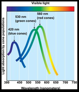
Cones
Allow for detailed color vision; densest in the center of the retina (fovea centralis).
Three types of cones, each sensitive to different wavelengths (red, green, blue).
Color blindness: Lack of one or more cone types.
Blind Spot
The optic disc is the region where the optic nerve exits the eye; no photoreceptors are present, creating a blind spot.
15-4 The Lens and Eye Chambers
Lens
Biconvex, crystal-like structure held in place by the ciliary zonule.
Focuses light on the retina.
Cataracts: Lens becomes hard and opaque with age, leading to vision loss.
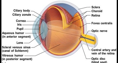
Eye Chambers
Anterior (aqueous) segment: Contains aqueous humor, maintains intraocular pressure, and provides nutrients.
Posterior (vitreous) segment: Contains vitreous humor, helps maintain eye shape and pressure.
15-5 Pathway of Light Through the Eye
Light passes through the conjunctiva, cornea, aqueous humor, pupil, lens, vitreous humor, and finally reaches the retina, where rods and cones are activated. The signal is transmitted via bipolar and ganglion cells to the optic nerve.

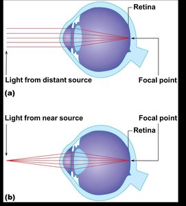
15-6 Image Formation on the Retina
Light is refracted by the cornea, aqueous humor, lens, and vitreous humor.
The eye is set for distance vision; accommodation allows the lens to change shape for near vision.
The image formed is real, reversed left to right, upside down, and smaller than the object. The brain corrects the image orientation.

15-7 Visual Fields and Visual Pathways
Optic chiasma: Site where fibers from the medial side of each eye cross to the opposite side of the brain.
Optic tracts: Contain fibers from both eyes, providing depth perception.
Pathway: Optic nerve → Optic chiasma → Optic tract → Thalamus → Optic radiation → Visual cortex in occipital lobe.
Hemianopia: Loss of the same side of the visual field in both eyes due to damage to one side of the visual cortex.
15-8 Eye Reflexes
Photopupillary reflex: Pupils constrict in bright light via the iris and ciliary muscles.
Accommodation pupillary reflex: Viewing close objects causes the lens to bulge and the eyes to converge medially.
15-9 Eye Terms and Disorders
Emmetropia: Normal vision; images focused correctly on the retina.
Myopia (nearsightedness): Distant objects are blurry; light focuses in front of the retina due to an elongated eyeball.
Hyperopia (farsightedness): Near objects are blurry; light focuses behind the retina due to a short eyeball or lazy lens.
Astigmatism: Blurry images due to unequal curvatures of the cornea or lens, causing light to focus as lines rather than points.
15-10 The Ear: Hearing and Equilibrium
Ear Anatomy
External ear: Auricle (pinna) and external acoustic meatus; involved in hearing only.
Middle ear (tympanic cavity): Air-filled cavity containing the tympanic membrane and ossicles (malleus, incus, stapes).
Inner ear (bony labyrinth): Contains the cochlea, vestibule, and semicircular canals; filled with perilymph and endolymph.
Ossicles and Sound Transmission
Ossicles transmit vibrations from the tympanic membrane to the oval window of the inner ear.
Pharyngotympanic (auditory) tube equalizes pressure between the middle ear and throat.
15-11 Organs of Equilibrium
Static Equilibrium
Maculae in the vestibule detect head position and linear acceleration.
Hair cells embedded in the otolithic membrane are bent by otoliths, triggering nerve impulses.
Dynamic Equilibrium
Crista ampullaris in the semicircular canals detects angular or rotary movements.
Movement of the cupula stimulates hair cells, sending signals to the cerebellum.
15-12 Organs of Hearing
Spiral organ of Corti: Located in the cochlear duct; contains hair cells (mechanoreceptors) for hearing.
Sound waves cause movement of the tectorial membrane, bending hair cells and generating nerve impulses.
15-13 Hearing and Equilibrium Deficits
Conduction deafness: Impaired transmission of sound through the external or middle ear (e.g., earwax, otitis media, otosclerosis).
Sensorineural deafness: Damage to hair cells, cochlear nerve, or auditory cortex (e.g., congenital defects, trauma, loud sounds).
Ménière’s syndrome: Progressive deafness, vertigo, nausea, and tinnitus due to excessive fluid pressure in the inner ear.
15-14 Chemical Senses: Taste and Smell
Olfaction (Smell)
Olfactory receptors are located in the roof of the nasal cavity; detect chemicals dissolved in mucus.
Impulses are transmitted via the olfactory nerve to the cerebrum for interpretation.
Taste (Gustation)
Taste buds are found on the tongue, soft palate, cheeks, and epiglottis.
Located on papillae (fungiform and vallate).
Gustatory cells possess microvilli (gustatory hairs) that detect chemicals dissolved in saliva.
Impulses are carried by the facial, glossopharyngeal, and vagus nerves to the gustatory cortex.
Five Basic Taste Sensations
Taste | Stimulus |
|---|---|
Sweet | Sugars, saccharine, some amino acids |
Sour | H+ ions (acids) |
Bitter | Alkaloids |
Salty | Metal ions |
Umami | Glutamate, "beefy" taste |
15-16 Developmental Aspects of the Special Senses
Special sense organs form early in embryonic development.
Maternal infections can cause visual and auditory abnormalities.
Vision requires significant postnatal development; infants are initially farsighted and lack depth perception.
Age-related changes include presbyopia (decreased lens elasticity), glaucoma, cataracts, and arteriosclerosis of ocular vessels.
Presbycusis (age-related hearing loss) may result from ossicle fusion (otosclerosis).
Taste and smell are most acute at birth and decline after age 40.
