 Back
BackThe Special Senses: Structure, Function, and Pathways
Study Guide - Smart Notes
 Tailored notes based on your materials, expanded with key definitions, examples, and context.
The Special Senses
General vs. Special Senses
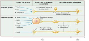
The human sensory system is divided into general senses and special senses. General senses include touch, pain, and temperature, which are detected by simple receptors distributed throughout the body. Special senses—taste, smell, vision, hearing, and equilibrium—are detected by specialized receptor cells located in specific organs.
General Senses: Detected by receptive endings of sensory neurons; include touch, pain, and temperature.
Special Senses: Detected by specialized receptor cells or structures; include taste, smell, vision, hearing, and equilibrium.
Location of Sensory Nerves: General senses use axons of spinal or cranial nerves, while special senses use axons of cranial nerves only.
Olfaction (Sense of Smell)
Anatomy of the Olfactory Epithelium
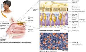
The olfactory epithelium is located in the superior region of the nasal cavity and contains several cell types that contribute to the sense of smell.
Olfactory Receptor Cells: Bipolar neurons with cilia (olfactory hairs) that detect odorants.
Supporting Cells: Provide metabolic and physical support to receptor cells.
Basal Cells: Stem cells that replace olfactory receptor cells.
Olfactory Glands: Produce mucus to dissolve odorants for detection.
Olfactory Transmission Pathway
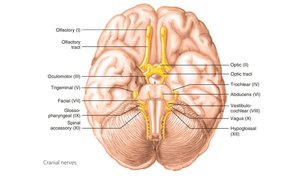
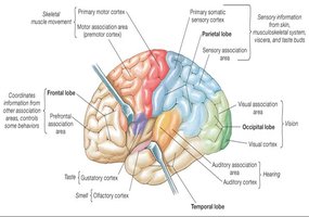
Odorant molecules bind to receptors on olfactory cilia, generating action potentials that travel through the olfactory nerve (cranial nerve I) to the olfactory bulb. From there, signals are relayed via the olfactory tract to the primary olfactory cortex in the brain.
Olfactory Nerve (I): Transmits signals from the nasal cavity to the olfactory bulb.
Olfactory Bulb: Processes and relays olfactory information.
Olfactory Tract: Carries processed signals to the brain.
Gustation (Sense of Taste)
Papillae of the Tongue
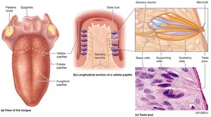
The tongue is covered with papillae, which are projections that house taste buds or provide texture sensation.
Vallate (Circumvallate) Papillae: Largest, dome-shaped, contain hundreds of taste buds.
Fungiform Papillae: Mushroom-shaped, contain a few taste buds each.
Foliate Papillae: Ridges on the side of the tongue, taste buds present mainly in childhood.
Filiform Papillae: Long, thin, do not contain taste buds; detect texture and temperature.
Anatomy of a Taste Bud
Taste buds are sensory organs for taste, located mainly on papillae. Each taste bud contains gustatory cells, supporting cells, and basal cells.
Gustatory Cells: Detect tastants and synapse with sensory neurons.
Supporting Cells: Provide structural support.
Basal Cells: Replace gustatory cells as they wear out.
Taste Sensations
There are five primary taste sensations, each associated with specific molecules:
Sweet: Simple sugars (e.g., glucose, fructose).
Sour: Hydrogen ions (e.g., in citric acid).
Salty: Metal ions (e.g., sodium, potassium).
Bitter: Various compounds, often toxic (protective function).
Umami: Glutamate and other amino acids (savory taste).
Gustatory Transduction
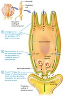
Gustatory transduction is the process by which tastants are converted into electrical signals in gustatory cells. Tastants bind to receptors, causing ion channels to open, leading to depolarization and neurotransmitter release.
The Gustatory Pathway
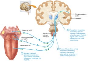
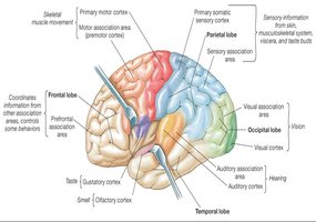
Taste signals are transmitted from the tongue to the brain via cranial nerves VII (facial), IX (glossopharyngeal), and X (vagus). These signals synapse in the solitary nucleus of the medulla, then travel to the thalamus and finally to the primary gustatory cortex in the insular lobe.
Additional info:
Special senses are localized to specific organs and have dedicated neural pathways, making them distinct from general senses.
Understanding the anatomy and physiology of special senses is crucial for diagnosing sensory disorders and understanding human interaction with the environment.
