 Back
BackThe Special Senses: Vision, Equilibrium, and Hearing
Study Guide - Smart Notes
 Tailored notes based on your materials, expanded with key definitions, examples, and context.
The Special Senses
Introduction to the Special Senses
The special senses are specialized sensory systems that provide information about the external environment. These include olfaction (smell), gustation (taste), vision, equilibrium (balance), and hearing. This chapter focuses on the structure and function of the organs responsible for vision and hearing, as well as the mechanisms underlying these senses.
Vision: The Eye
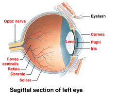
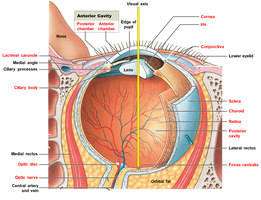
Anatomy of the Eyeball
The eyeball is a hollow spheroid filled with fluid and situated within the orbit. It is protected and cushioned by orbital fat and surrounded by extrinsic eye muscles, the lacrimal gland, cranial nerves, and blood vessels.
Two interior cavities:
Anterior cavity: Contains aqueous humor; subdivided into the anterior chamber (between cornea and iris) and posterior chamber (between iris and lens).
Posterior cavity: Contains the vitreous body (gelatinous substance).
Layers of the Eye
Fibrous Layer:
Sclera: The "white of the eye," composed of dense fibrous connective tissue, provides structure and protection.
Cornea: Transparent anterior portion, continuous with the sclera, allows light to enter the eye. It is avascular and has limited repair capacity.
Vascular Layer (Uvea):
Iris: Pigmented ring containing blood vessels, melanocytes, and pupillary muscles. Controls the diameter of the pupil, regulating light entry.
Ciliary Body: Smooth muscle and fibers that hold the lens in place and regulate its shape.
Choroid: Vascular layer posterior to the ora serrata, supplies oxygen and nutrients to the retina.
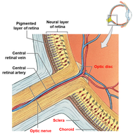
Inner Layer (Retina):
Pigmented Layer: Absorbs light and prevents reflection within the eye.
Neural Layer: Contains photoreceptors (rods and cones), supporting cells, and neurons.
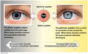
The Iris and Pupillary Muscles
The iris contains two sets of smooth muscles: the sphincter pupillae (constricts the pupil) and the dilator pupillae (dilates the pupil). These muscles regulate the amount of light entering the eye in response to autonomic nervous system stimulation.
Accessory and Internal Structures
Lens: Transparent, biconvex, flexible disc that focuses light onto the retina. Held in place by the ciliary body and surrounded by a dense elastic capsule.
Aqueous Humor: Watery fluid in the anterior cavity, provides nutrients, removes waste, and maintains intraocular pressure.
Vitreous Body: Gelatinous mass in the posterior cavity, stabilizes eye shape.
The Retina and Photoreceptors
Rods: Highly sensitive to light, enable vision in low-light conditions, do not detect color, more numerous in the peripheral retina.
Cones: Responsible for color vision and sharp visual acuity, concentrated in the macula and especially the fovea centralis.
Optic Disc: Site where the optic nerve exits the eye; lacks photoreceptors and is known as the "blind spot."
Circulation of Aqueous Humor and Intraocular Pressure
Aqueous humor is produced by the ciliary body, circulates through the posterior and anterior chambers, and is reabsorbed via the scleral venous sinus. Proper drainage is essential to maintain intraocular pressure; impaired drainage can lead to glaucoma, which may damage the optic nerve.
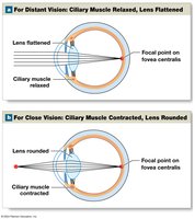
Refraction and Focusing of Light
Light is refracted as it passes through the cornea and lens, focusing the image on the retina. The lens changes shape (accommodation) to focus on objects at different distances:
Rounder lens: Focuses on near objects.
Flatter lens: Focuses on distant objects.
Common Refractive Problems
Astigmatism: Irregular curvature of the cornea or lens causes distorted images.
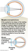
Myopia (Nearsightedness): Image focuses in front of the retina; corrected with diverging lenses.
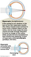
Hyperopia (Farsightedness): Image focuses behind the retina; corrected with converging lenses.
Equilibrium and Hearing: The Ear
Anatomy of the Ear
The ear is divided into three main regions: the external ear, middle ear, and internal ear. Each region has specialized structures for the detection and transmission of sound and equilibrium sensations.
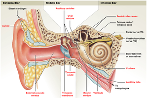
External Ear
Auricle (Pinna): Collects and directs sound waves into the external acoustic meatus.
External Acoustic Meatus: Canal that channels sound to the tympanic membrane.
Tympanic Membrane (Eardrum): Vibrates in response to sound waves, transmitting vibrations to the middle ear.
Middle Ear (Tympanic Cavity)
Auditory Tube (Eustachian Tube): Equalizes pressure between the middle ear and nasopharynx.
Auditory Ossicles:
Malleus (Hammer): Attached to the tympanic membrane.
Incus (Anvil): Connects malleus to stapes.
Stapes (Stirrup): Transmits vibrations to the oval window of the internal ear.
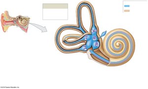
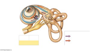
Internal Ear (Labyrinth)
Vestibule: Contains the saccule and utricle, which detect gravity and linear acceleration.
Semicircular Canals: Contain semicircular ducts that detect rotational movements.
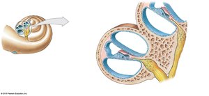
Cochlea: Spiral-shaped structure containing the cochlear duct, responsible for hearing.
Membranous and Bony Labyrinths: The membranous labyrinth is filled with endolymph, while the bony labyrinth contains perilymph.
Equilibrium
Equilibrium sensations are detected by hair cells in the vestibular complex (vestibule and semicircular canals). These cells respond to changes in head position and movement, providing information about balance and spatial orientation.
Hearing
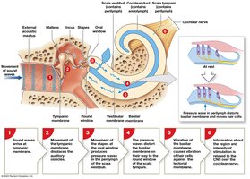
Hearing involves the conversion of sound waves into mechanical vibrations, which are then transmitted through the auditory ossicles to the internal ear. In the cochlea, these vibrations are converted into pressure waves in the fluid, stimulating hair cells in the spiral organ (organ of Corti).
Cochlear Duct (Scala Media): Contains endolymph and the spiral organ.
Scala Vestibuli and Scala Tympani: Chambers filled with perilymph, located above and below the cochlear duct, respectively.
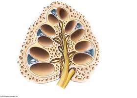
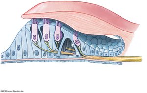
The Spiral Organ (Organ of Corti)
The spiral organ rests on the basilar membrane within the cochlear duct. It contains rows of hair cells whose stereocilia contact the overlying tectorial membrane. Movement of the basilar membrane causes the stereocilia to bend, opening ion channels and initiating nerve impulses.

Steps in the Process of Hearing
Sound waves vibrate the tympanic membrane.
Vibrations are transmitted and amplified by the auditory ossicles.
Stapes movement at the oval window creates pressure waves in the perilymph of the scala vestibuli.
Pressure waves distort the basilar membrane, stimulating hair cells in the spiral organ.
Hair cell depolarization leads to nerve impulses transmitted via the cochlear nerve (part of cranial nerve VIII) to the brain.
Auditory Discrimination and Age-Related Changes
The human ear can detect a wide range of sound intensities and frequencies.
With age, hearing sensitivity decreases due to stiffening of the tympanic membrane, ossicle articulations, and possible ossification of the round window.
Summary Table: Main Structures and Functions of the Eye and Ear
Structure | Location | Function |
|---|---|---|
Cornea | Anterior eye | Refracts light, protects eye |
Lens | Behind iris | Focuses light on retina |
Retina | Inner layer of eye | Contains photoreceptors for vision |
Auricle | External ear | Collects sound waves |
Tympanic membrane | Between external and middle ear | Transmits sound vibrations |
Cochlea | Internal ear | Detects sound |
Semicircular canals | Internal ear | Detect rotational movement (equilibrium) |
Additional info: This guide expands on the provided lecture slides with definitions, examples, and a summary table for clarity and exam preparation.
