 Back
BackThe Spinal Cord and Spinal Nerves: Structure, Function, and Clinical Relevance
Study Guide - Smart Notes
 Tailored notes based on your materials, expanded with key definitions, examples, and context.
The Spinal Cord and Spinal Nerves
Overview of the Spinal Cord
The spinal cord, together with the brain, forms the central nervous system (CNS). It serves as a major pathway for transmitting sensory and motor information between the body and the brain, and is also responsible for reflex actions and integration of nerve impulses.
Functions: Reflexes, integration of nerve impulses, and conduction of sensory/motor information.
Length: Approximately 16–18 inches in adults; ends at L2 in adults and L4 in newborns.
Enlargements: Cervical and lumbar enlargements correspond to the origin of nerves for the upper and lower limbs.
Protection of the Spinal Cord
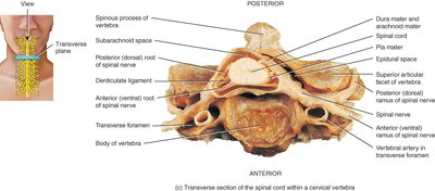
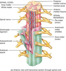
The spinal cord is protected by bony vertebrae, connective tissue coverings called meninges, and a cushion of cerebrospinal fluid (CSF).
Meninges: Three continuous layers—dura mater (outer), arachnoid mater (middle), and pia mater (inner).
Epidural space: Contains fat and blood vessels between vertebrae and dura mater.
Subarachnoid space: Located between arachnoid and pia mater; contains CSF.
Clinical Applications
Meningitis: Inflammation of the meninges, often due to infection.
Spinal Tap (Lumbar Puncture): Removal of CSF from the subarachnoid space for diagnostic or therapeutic purposes.
External and Inferior Anatomy of the Spinal Cord
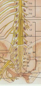
Conus medullaris: Cone-shaped end of the spinal cord.
Filum terminale: Thread-like extension of pia mater that stabilizes the spinal cord.
Cauda equina: Bundle of dorsal and ventral roots of the lowest spinal nerves, resembling a horse's tail.
Spinal Nerves
Classification and Structure
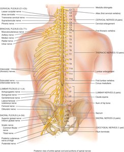
There are 31 pairs of spinal nerves, named and numbered according to the region of the spinal cord from which they emerge. Each spinal nerve is a mixed nerve, carrying both sensory and motor fibers.
8 pairs of cervical nerves (C1–C8)
12 pairs of thoracic nerves (T1–T12)
5 pairs of lumbar nerves (L1–L5)
5 pairs of sacral nerves (S1–S5)
1 pair of coccygeal nerves (Co1)

Connective Tissue Coverings of Spinal Nerves
Endoneurium: Surrounds individual axons.
Perineurium: Surrounds bundles of axons (fascicles).
Epineurium: Surrounds the entire nerve.
Branching and Plexuses
After emerging from the spinal cord, each spinal nerve branches into dorsal and ventral rami. The ventral rami (except in the thoracic region) form nerve plexuses—networks of nerves that supply specific body regions.
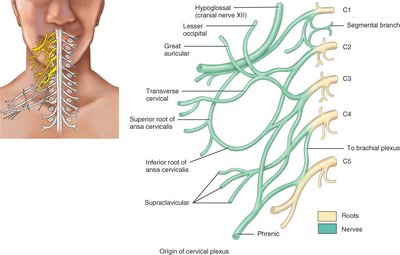
Cervical plexus (C1–C5): Supplies head, neck, shoulders; includes the phrenic nerve (C3–C5) to the diaphragm.
Brachial plexus (C5–T1): Supplies upper limbs.
Lumbar plexus (L1–L4): Supplies anterior and medial thigh.
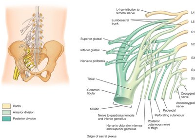
Sacral plexus (L4–S4): Supplies buttocks, perineum, lower limbs; includes the sciatic nerve.
Internal Anatomy of the Spinal Cord
Gray and White Matter
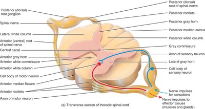
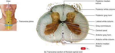
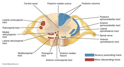
The spinal cord is organized into gray matter (shaped like an H or butterfly) and white matter. The gray matter contains neuron cell bodies and dendrites, while the white matter consists of myelinated axons organized into tracts.
Gray horns: Dorsal (posterior), ventral (anterior), and lateral (only in thoracic region).
White columns: Anterior, lateral, and posterior columns contain ascending (sensory) and descending (motor) tracts.
Central canal: Contains CSF.
Spinal Cord Tracts
Tracts are bundles of axons in the white matter that carry specific types of information.
Sensory (ascending) tracts: Carry sensory information to the brain (e.g., spinothalamic tract for pain and temperature, posterior columns for proprioception and touch).
Motor (descending) tracts: Carry motor commands from the brain to effectors (e.g., corticospinal tracts for voluntary movement).
Spinal Cord Physiology
Functions of the Spinal Cord
White matter tracts: Serve as highways for nerve impulse conduction to and from the brain.
Gray matter: Integrates incoming and outgoing information, serving as the center for reflexes.
Reflexes and Reflex Arcs
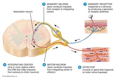
A reflex is a fast, predictable, automatic response to a stimulus, helping maintain homeostasis. Reflexes can be spinal or cranial, somatic or autonomic. The reflex arc is the specific pathway followed by nerve impulses during a reflex.
Components of a reflex arc: Receptor, sensory neuron, integrating center, motor neuron, effector.
Clinical Considerations: Reflex Testing
Stretch reflex (e.g., patellar reflex): Monosynaptic, ipsilateral; helps prevent muscle overstretching.
Plantar reflex: Stroking the sole of the foot; abnormal response in adults (Babinski sign) may indicate CNS damage.
Dermatomes and Clinical Relevance
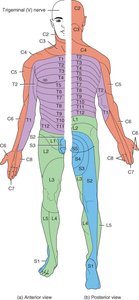
Dermatomes
A dermatome is an area of skin supplied by sensory fibers from a single spinal nerve. Mapping dermatomes helps clinicians diagnose the level of spinal cord or nerve damage.
All spinal nerves except C1 innervate specific skin segments.
Damage to a spinal nerve or cord segment can be detected by patterns of numbness.
Spinal Cord Injury and Disease
Spinal cord transection: Complete severing of the cord results in loss of sensation and motor control below the injury.
Shingles: Infection of peripheral nerves by the varicella-zoster virus, causing pain and blisters along a dermatome.
