 Back
BackThe Spinal Cord and Spinal Nerves: Structure, Function, and Pathways
Study Guide - Smart Notes
 Tailored notes based on your materials, expanded with key definitions, examples, and context.
Spinal Cord and Spinal Nerves
Principal Functions of the Spinal Cord
The spinal cord is a critical component of the central nervous system, serving as a conduit for information between the brain and the rest of the body. It also coordinates reflexes andprocesses sensory and motor information.
Conduction: Transmits sensory and motor signals between the brain and peripheral nerves.
Integration: Processes and integrates incoming sensory information and outgoing motor commands.
Reflexes: Mediates rapid, involuntary responses to stimuli (spinal reflexes).
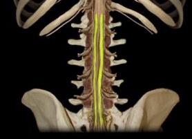
Gross Anatomy of the Spinal Cord
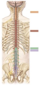
The spinal cord is divided into five regions, each associated with a group of spinal nerves. It is protected by vertebrae, meninges, and cerebrospinal fluid.
Cervical: Continuous with the medulla oblongata; contains neurons for cervical spinal nerves.
Thoracic: Contains neurons for thoracic spinal nerves.
Lumbar: Short segment with neurons for lumbar spinal nerves.
Sacral: Contains neurons for sacral spinal nerves.
Coccygeal: Most inferior tip of the cord.
Cervical and Lumbar Enlargements: Regions with increased numbers of neurons for limb innervation.
Spinal Nerves: Identification and Structure
Spinal nerves are mixed nerves carrying both sensory and motor fibers. There are 31 pairs, classified by region:
8 cervical
12 thoracic
5 lumbar
5 sacral
1 coccygeal
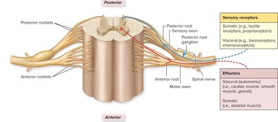
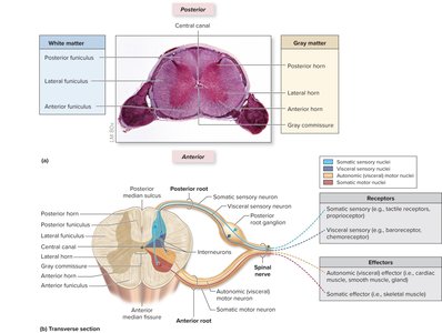
Spinal Roots and Nerve Anatomy
Each spinal nerve is formed by the union of an anterior (motor) root and a posterior (sensory) root. The posterior root contains a ganglion housing sensory neuron cell bodies.

Protection and Support of the Spinal Cord
The spinal cord is protected by the vertebral column, meninges (dura mater, arachnoid mater, pia mater), and cerebrospinal fluid. The meninges provide structural support and a barrier against pathogens.
Epidural space: Contains fat and blood vessels.
Dura mater: Tough outer layer.
Subdural space: Potential space between dura and arachnoid.
Arachnoid mater: Middle, web-like layer.
Subarachnoid space: Contains cerebrospinal fluid (CSF).
Pia mater: Delicate inner layer adhering to the spinal cord.
Clinical Application: Lumbar Puncture
A lumbar puncture is a procedure to obtain CSF for diagnostic purposes. The needle is inserted below the level of the spinal cord (usually between L3 and L4) to avoid injury.
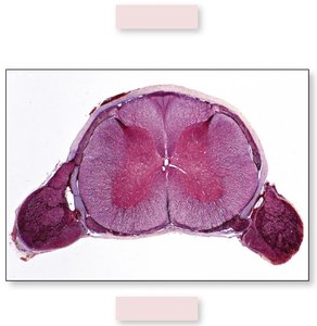
Internal Anatomy: Gray and White Matter
The spinal cord's cross-section reveals a central H-shaped region of gray matter (neuron cell bodies, dendrites, unmyelinated axons) surrounded by white matter (myelinated axons).
Gray Matter: Divided into anterior, lateral, and posterior horns.
White Matter: Organized into posterior, lateral, and anterior funiculi containing ascending (sensory) and descending (motor) tracts.
Nervous System Pathways: Ascending and Descending Tracts
Nervous system pathways are organized as sensory (ascending) or motor (descending) tracts. Most pathways are paired and decussate (cross the midline), resulting in contralateral control.
Sensory Pathways: Carry information from receptors to the brain.
Motor Pathways: Transmit commands from the brain to effectors (muscles/glands).
Sensory Pathways
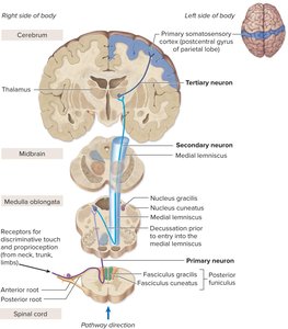
Posterior Funiculus–Medial Lemniscal Pathway: Discriminative touch, proprioception, and visceral pain. Involves three neurons (primary, secondary, tertiary).
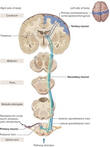
Anterolateral (Spinothalamic) Pathway: Crude touch, pressure, pain, temperature. Also a three-neuron chain.
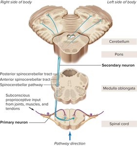
Spinocerebellar Pathway: Proprioceptive information to the cerebellum; uses two neurons.

Motor Pathways
Direct (Pyramidal) Pathway: Controls voluntary movements via corticospinal tracts (lateral and anterior).
Indirect Pathways: Regulate muscle tone, posture, and reflexes (rubrospinal, reticulospinal, tectospinal, vestibulospinal tracts).
Spinal Nerves: Branches and Dermatomes
After exiting the vertebral column, each spinal nerve splits into branches (rami):
Posterior (dorsal) ramus: Innervates muscles and skin of the back.
Anterior (ventral) ramus: Innervates anterior/lateral trunk and limbs; forms plexuses.
Rami communicantes: Connect spinal nerves to the sympathetic trunk ganglia.
Dermatomes are regions of skin supplied by a single spinal nerve. Dermatome maps are used clinically to localize nerve damage and understand referred pain patterns.
Reflexes and Reflex Arcs
A reflex is a rapid, involuntary, and stereotyped response to a stimulus. Reflex arcs consist of five components:
Receptor
Afferent (sensory) neuron
Integrating center (spinal cord)
Efferent (motor) neuron
Effector (muscle or gland)
Somatic reflexes involve skeletal muscles and are essential for posture and protection.
Clinical Considerations: Spinal Cord Injury and Disease
Spinal Cord Injury: Can result in paralysis and loss of sensation below the injury site. Prompt treatment with steroids and antibiotics can improve outcomes. Research into neural stem cells offers hope for future therapies.
Shingles (Herpes Zoster): Reactivation of varicella-zoster virus in posterior root ganglia causes painful dermatomal rash.
Summary Table: Spinal Cord Regions and Functions
Region | Associated Nerves | Main Function |
|---|---|---|
Cervical | C1–C8 | Neck, upper limb innervation |
Thoracic | T1–T12 | Trunk, thoracic organs |
Lumbar | L1–L5 | Lower limb innervation |
Sacral | S1–S5 | Pelvic organs, lower limbs |
Coccygeal | Co1 | Small region near coccyx |
