 Back
The Spinal Cord, Spinal Nerves, and Spinal Reflexes: Structure and Function
Study Guide - Smart Notes
 Tailored notes based on your materials, expanded with key definitions, examples, and context.
The Nervous System and Reflexes
Reflexes and Spinal Reflexes
Reflexes are quick, automatic nerve responses triggered by specific stimuli. Spinal reflexes are controlled by the spinal cord alone, without input from the brain. These reflexes are essential for rapid responses to potentially harmful stimuli, such as dropping a hot pan or stepping on something sharp.
The Structure and Function of the Spinal Cord
Overview of the Spinal Cord
The spinal cord is located within the vertebral column and is surrounded by protective membranes called meninges. It serves as a major pathway for carrying sensory and motor information between the body and the brain and gives rise to spinal nerves that innervate various body regions.
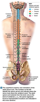
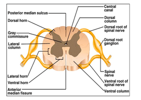
Spinal Cord Anatomy: Cross-Sectional Structure
The spinal cord contains both gray matter (cell bodies, neuroglia, and unmyelinated axons) and white matter (myelinated axons). Key anatomical features include:
Posterior median sulcus (shallow groove on the posterior side)
Anterior median fissure (deeper groove on the anterior side)
Central canal (contains cerebrospinal fluid, CSF)
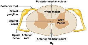
Spinal Cord Roots and Ganglia
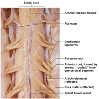
Each spinal nerve is formed by the junction of two roots:
Anterior (ventral) root: Contains axons of motor neurons
Posterior (dorsal) root: Contains axons of sensory neurons
Spinal ganglia (dorsal root ganglia): Contain cell bodies of sensory neurons
The Spinal Meninges
The spinal cord is protected by three specialized membranes called meninges:
Dura mater: Outermost, tough layer; continuous with cranial dura mater
Arachnoid mater: Middle layer; contains the subarachnoid space filled with CSF
Pia mater: Innermost, delicate layer; firmly attached to the spinal cord surface
These layers protect the spinal cord and provide structural stability. Infections of the meninges are called meningitis.
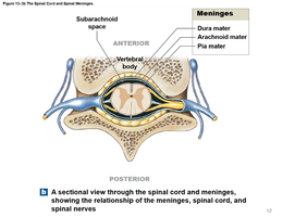
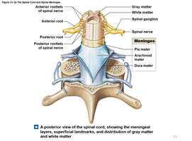
Spaces Associated with the Meninges
Epidural space: Between vertebrae and dura mater; contains loose connective and adipose tissue
Subdural space: Potential space deep to dura mater
Subarachnoid space: Filled with CSF; site for lumbar puncture (spinal tap)
Pia Mater and Denticulate Ligaments
The pia mater is attached to the spinal cord and contains blood vessels. Denticulate ligaments anchor the pia mater to the dura mater, preventing lateral movement of the spinal cord.
Gray and White Matter Organization
Gray Matter
Gray matter consists of neuron cell bodies, neuroglia, and unmyelinated axons. It is organized into:
Dorsal horn: Sensory processing
Lateral horn: Autonomic motor neurons (in thoracic and upper lumbar regions)
Ventral horn: Somatic motor neurons
White Matter
White matter contains myelinated axons organized into columns (funiculi):
Dorsal column
Lateral column
Ventral column
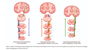
Tracts within these columns relay information:
Ascending tracts: Carry sensory information to the brain
Descending tracts: Carry motor commands from the brain
Spinal Nerves and Plexuses
Structure of Spinal Nerves
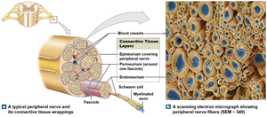
Spinal nerves are mixed nerves containing both sensory (afferent) and motor (efferent) fibers. Each nerve is surrounded by three connective tissue layers:
Epineurium: Outermost, dense collagen fibers
Perineurium: Middle layer, divides nerve into fascicles (axon bundles)
Endoneurium: Innermost, surrounds individual axons
Dermatomes and Peripheral Neuropathies
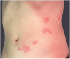
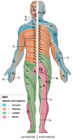
A dermatome is a specific bilateral region of skin supplied by a single pair of spinal nerves. Damage or disease affecting spinal nerves can lead to peripheral neuropathies, resulting in regional losses of neural function. Shingles is a viral infection that causes a rash along the affected dermatome.
Nerve Plexuses
Nerve plexuses are complex, interwoven networks of nerve fibers formed from the anterior rami of adjacent spinal nerves. They allow multiple spinal nerves to supply the same structures. The four major plexuses are:
Cervical plexus
Brachial plexus
Lumbar plexus
Sacral plexus
Neural Reflexes
Reflex Arcs
A reflex arc is the basic functional unit of the nervous system, producing rapid, automatic responses to specific stimuli. The five components of a reflex arc are:
Sensory receptor
Sensory neuron
Information processing in the CNS (often involving interneurons)
Motor neuron
Effector (muscle or gland)
Classification of Reflexes
Development:
Innate reflexes: Genetically programmed, present at birth (e.g., withdrawal reflex)
Acquired reflexes: Learned through experience (e.g., braking a car in an emergency)
Motor response:
Somatic reflexes: Control skeletal muscle contractions (e.g., patellar reflex)
Visceral (autonomic) reflexes: Control smooth muscle, cardiac muscle, or glands
Summary Table: Spinal Cord and Spinal Nerve Organization
Structure | Function |
|---|---|
Gray Matter | Cell bodies, neuroglia, unmyelinated axons; processing and integration |
White Matter | Myelinated axons; transmission of signals (ascending and descending tracts) |
Spinal Nerves | Mixed nerves; connect CNS to body regions |
Meninges | Protect and stabilize the spinal cord |
Dermatome | Skin region supplied by a single spinal nerve |
Nerve Plexus | Network of nerves supplying limbs and body wall |
