Back
The Spinal Cord, Spinal Nerves, and Spinal Reflexes: Structured Study Notes
Study Guide - Smart Notes
 Tailored notes based on your materials, expanded with key definitions, examples, and context.
The Nervous System: Overview and Organization
Central and Peripheral Nervous System
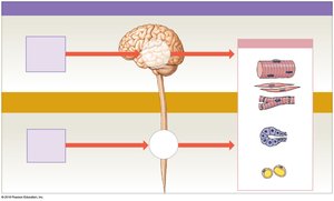
The nervous system is divided into the central nervous system (CNS) and the peripheral nervous system (PNS). The CNS consists of the brain and spinal cord, which serve as processing centers for sensory input and motor output. The PNS includes cranial and spinal nerves that connect the CNS to the rest of the body, facilitating communication between sensory receptors and effectors such as muscles and glands.
CNS: Brain and spinal cord; responsible for processing and integrating information.
PNS: Cranial nerves (arising from the brain) and spinal nerves (arising from the spinal cord); responsible for transmitting sensory input and motor commands.
Reflexes: Quick, automatic responses to specific stimuli, often mediated by the spinal cord without brain involvement.
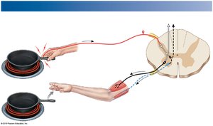
Example: Dropping a hot pan is a spinal reflex; the hand releases the pan before pain is consciously perceived.
The Spinal Cord: Structure and Function
Gross Anatomy of the Spinal Cord
The spinal cord is a cylindrical structure housed within the vertebral column and protected by meninges. It serves as a major pathway for transmitting information between the brain and the rest of the body.
Length: Approximately 45 cm (18 in.) long and 14 mm (1/2 in.) wide.
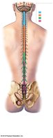
Regions: Cervical, thoracic, lumbar, and sacral.
Segments: 31 segments, each giving rise to a pair of spinal nerves.
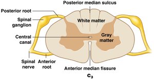
Grooves: Posterior median sulcus (shallow, posterior), anterior median fissure (deeper, anterior).
Central canal: Contains cerebrospinal fluid (CSF).
Enlargements and Distal End of the Spinal Cord
Cervical enlargement: Supplies nerves to the shoulder and upper limbs.
Lumbosacral enlargement: Supplies nerves to the pelvis and lower limbs.
Conus medullaris: Tapered end of the spinal cord below the lumbar enlargement.
Cauda equina: Bundle of nerve roots extending below the conus medullaris.
Filum terminale: Thin thread of fibrous tissue anchoring the spinal cord to the coccyx.
Spinal Roots, Ganglia, and Nerves
Ventral (anterior) root: Contains motor neuron axons.
Dorsal (posterior) root: Contains sensory neuron axons; associated with dorsal root ganglia (cell bodies of sensory neurons).
Spinal nerves: Formed by the union of dorsal and ventral roots; mixed nerves containing both sensory and motor fibers.
Rami: Branches of spinal nerves; posterior ramus supplies the back, anterior ramus supplies the body wall and limbs.
Cross-Sectional Anatomy of the Spinal Cord
The spinal cord's cross-section reveals the arrangement of gray and white matter, as well as the roots and ganglia associated with spinal nerves.
Gray matter: Contains neuron cell bodies, neuroglia, and unmyelinated axons; organized into horns (anterior, posterior, lateral).
White matter: Contains myelinated and unmyelinated axons; organized into columns (anterior, posterior, lateral).
Central canal: Runs through the center, containing CSF.
The Spinal Meninges
Protective Membranes
The spinal cord is protected by three layers of connective tissue called meninges:
Dura mater: Outermost, tough layer; continuous with cranial dura mater.
Arachnoid mater: Middle, weblike layer; contains the subarachnoid space filled with CSF.
Pia mater: Innermost, delicate layer; adheres to the spinal cord surface and contains blood vessels.
Meningitis is an infection of the meninges, which can be viral or bacterial.
Gray Matter and White Matter Organization
Functional Organization of Gray Matter
Nuclei: Collections of neuron cell bodies in the CNS.
Posterior horns: Somatic and visceral sensory nuclei.
Anterior horns: Somatic motor nuclei.
Lateral horns: Visceral motor nuclei (thoracic and lumbar regions).
Gray commissures: Bands of gray matter around the central canal, allowing communication between sides.
Functional Organization of White Matter
Columns: Posterior, anterior, and lateral white columns.
Tracts: Bundles of axons relaying the same type of information in the same direction.
Ascending tracts: Carry sensory information to the brain.
Descending tracts: Carry motor commands from the brain.
Spinal Nerves and Plexuses
Connective Tissue Layers of Spinal Nerves
Epineurium: Outermost layer, dense collagen fibers.
Perineurium: Middle layer, surrounds fascicles (bundles of axons).
Endoneurium: Innermost layer, surrounds individual axons.
Dermatomes and Peripheral Neuropathies
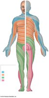
Dermatome: Specific region of skin supplied by a single pair of spinal nerves.
Peripheral neuropathies: Regional losses of neural function, often due to trauma or disease.
Shingles: Viral infection causing a rash along a dermatome.

Nerve Plexuses
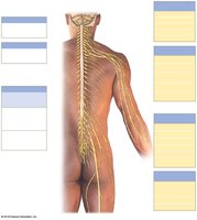
Plexus: Complex network of nerve fibers formed from anterior rami of adjacent spinal nerves.
Major plexuses: Cervical, brachial, lumbar, sacral.
Function: Allows multiple spinal nerves to supply the same structures, providing redundancy and flexibility.
Neuronal Pools and Neural Circuits
Patterns of Neural Circuits
Divergence: Spreads information from one neuron to many.
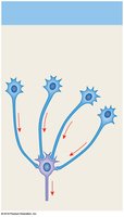
Convergence: Multiple neurons synapse on a single neuron.
Serial processing: Information moves sequentially through a series of neurons.
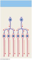
Parallel processing: Several neurons process the same information simultaneously.
Reverberation: Positive feedback loop that maintains activity until inhibited.
Reflexes and Reflex Arcs
Neural Reflexes
Definition: Rapid, automatic responses to specific stimuli; basic units of neural function.
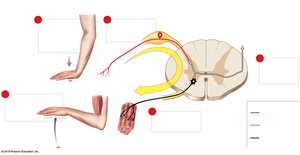
Reflex arc components: Sensory receptor, sensory neuron, integration center (CNS), motor neuron, effector.
Classification of Reflexes
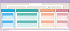
Criterion | Types |
|---|---|
Development | Innate (inborn) or acquired (learned) |
Response | Somatic (skeletal muscle) or visceral (autonomic) |
Complexity | Monosynaptic (one synapse) or polysynaptic (multiple synapses) |
Processing Site | Spinal or cranial |
Spinal Reflexes
Monosynaptic Reflexes: The Stretch Reflex
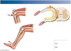
Function: Regulates skeletal muscle length; very rapid response.
Example: Patellar (knee-jerk) reflex.
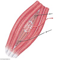
Muscle spindle: Receptor for stretch reflex; contains intrafusal fibers innervated by gamma motor neurons.
Polysynaptic Reflexes
Tendon reflex: Prevents excessive tension in muscles and tendons; involves Golgi tendon organs.
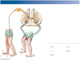
Withdrawal reflex: Moves body part away from painful stimulus; involves reciprocal inhibition.
Crossed extensor reflex: Coordinates actions on opposite sides of the body during withdrawal reflexes.
Brain Influence on Spinal Reflexes
Integration and Control
Facilitation: Brain can enhance reflexes by increasing excitatory input to spinal neurons.
Inhibition: Brain can suppress reflexes by increasing inhibitory input.
Examples: Voluntary movements such as walking or running involve both reflex and conscious control.
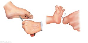
Plantar and Babinski Reflexes
Plantar reflex: Normal in adults; stroking the lateral sole causes toe curling.
Babinski reflex: Normal in infants; dorsiflexion of the big toe and fanning of other toes. In adults, indicates CNS damage.