 Back
BackThe Spinal Cord, Spinal Nerves, and Spinal Reflexes – Study Notes
Study Guide - Smart Notes
 Tailored notes based on your materials, expanded with key definitions, examples, and context.
Chapter 13: The Spinal Cord, Spinal Nerves, and Spinal Reflexes
Overview
This chapter explores the structure and function of the spinal cord, spinal nerves, and the mechanisms underlying spinal reflexes. Understanding these components is essential for grasping how the nervous system processes sensory input and coordinates motor output.
The Nervous System
Structural and Organizational Characteristics
Central Nervous System (CNS): Composed of the brain and spinal cord; serves as the main processing center for the entire nervous system.
Peripheral Nervous System (PNS): Includes cranial nerves and spinal nerves; responsible for transmitting sensory input to the CNS and carrying motor commands to effectors.
Reflexes: Quick, automatic responses to specific stimuli. Spinal reflexes are mediated by the spinal cord without brain involvement.
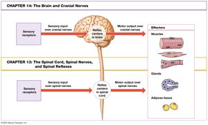
The Spinal Cord
Structure and Functions
The spinal cord is a cylindrical organ made of nervous tissue, protected by the vertebral column and meninges.
It transmits sensory and motor information between the brain and the rest of the body.
It is the origin of 31 pairs of spinal nerves, each corresponding to a segment of the spinal cord.
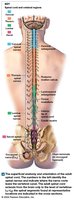
Gross Anatomy of the Adult Spinal Cord
Segments: 31 segments, each giving rise to a pair of spinal nerves.
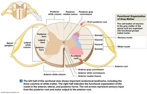
Bilateral Symmetry: The spinal cord is divided into left and right halves by the posterior median sulcus and anterior median fissure.
Central Canal: A passageway containing cerebrospinal fluid (CSF).
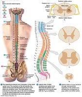
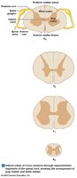
Spinal Roots and Ganglia
Anterior (Ventral) Root: Contains axons of motor neurons.
Posterior (Dorsal) Root: Contains axons of sensory neurons.
Spinal Ganglia (Dorsal Root Ganglia): Contain cell bodies of sensory neurons; located between vertebrae.
Spinal Nerves: Formed by the union of anterior and posterior roots; are mixed nerves (contain both sensory and motor fibers).
Spinal Meninges
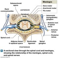
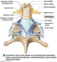
Specialized membranes that provide physical stability, shock absorption, and a pathway for blood vessels.
Dura Mater: Outermost, tough layer.
Arachnoid Mater: Middle, web-like layer.
Pia Mater: Innermost, delicate layer adhering to the spinal cord.
Continuous with cranial meninges.
Meningitis: Inflammation of the meninges due to infection.
Subarachnoid Space and Clinical Procedures
The subarachnoid space lies between the arachnoid and pia mater and contains CSF, which cushions the spinal cord and transports nutrients and wastes.
Lumbar Puncture (Spinal Tap): Procedure to withdraw CSF for diagnostic purposes.
Spinal Anesthesia: Local anesthetics administered into the subarachnoid space.
Gray Matter and White Matter
Organization and Function
Gray Matter: Contains neuron cell bodies, neuroglia, and unmyelinated axons; integrates information and initiates commands; located deep in the spinal cord.
White Matter: Contains myelinated and unmyelinated axons; transmits information; located superficially in the spinal cord.
Gray Matter Organization
Nuclei: Masses of gray matter in the CNS, organized into horns.
Posterior Horns: Somatic and visceral sensory nuclei (incoming information).
Anterior Horns: Somatic motor nuclei (outgoing commands).
Lateral Horns: Visceral motor nuclei (thoracic and lumbar segments).
Gray Commissure: Connects the two sides of gray matter around the central canal.
White Matter Organization
Divided into three columns: posterior, anterior, and lateral white columns.
Tracts: Bundles of axons carrying the same type of information in the same direction.
Ascending Tracts: Carry sensory information to the brain.
Descending Tracts: Carry motor commands from the brain to the spinal cord.
Spinal Nerves and Plexuses
Connective Tissue Layers of Spinal Nerves
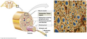
Epineurium: Outermost layer, dense collagen fibers.
Perineurium: Middle layer, divides nerve into fascicles (bundles of axons).
Endoneurium: Innermost layer, surrounds individual axons.
Peripheral Distribution and Clinical Relevance
Dermatome: A specific region of skin supplied by a single pair of spinal nerves; important for diagnosing nerve or spinal cord injuries.
Peripheral Neuropathies: Regional loss of sensory and motor function, often due to trauma or compression.
Shingles: Reactivation of varicella-zoster virus affecting dermatomes.
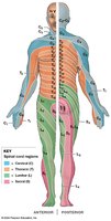
Nerve Plexuses
Nerve Plexus: A network of interwoven nerve fibers formed from the anterior rami of adjacent spinal nerves; allows multiple nerves to supply the same structures.
Four major plexuses: cervical, brachial, lumbar, and sacral.
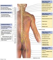
Cervical Plexus
Innervates the scalp, neck, and diaphragm.
Phrenic Nerve: Controls the diaphragm.
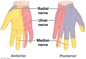
Brachial Plexus
Innervates the pectoral girdle, upper back, and upper limbs.
Musculocutaneous Nerve: Arm flexors, lateral forearm skin.
Radial Nerve: Arm and forearm extensors, posterolateral skin.
Median Nerve: Forearm and digital flexors, anterolateral hand skin.
Ulnar Nerve: Wrist and small digital muscle flexors, medial hand skin.
Lumbar and Sacral Plexuses
Lumbar Plexus: Femoral nerve (anterior thigh), obturator nerve (medial thigh).
Sacral Plexus: Superior/inferior gluteal nerves (gluteal muscles), posterior femoral cutaneous nerve (posterior thigh/leg), sciatic nerve (hamstrings, divides into fibular and tibial nerves).
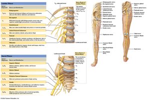
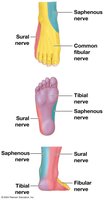
Sensory Innervation of the Foot
Supplied by the saphenous, sural, fibular, and tibial nerves.
Neuronal Pools
Functional Organization of Neurons
Sensory Neurons: Transmit information to the CNS.
Motor Neurons: Carry commands from the CNS to effectors.
Interneurons: Located within the CNS; interpret, plan, and coordinate signals.
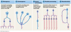
Patterns of Neural Circuits
Divergence: Spreads stimulation to multiple neurons or pools.
Convergence: Multiple sources provide input to a single neuron or pool.
Serial Processing: Neurons or pools process information sequentially.
Parallel Processing: Neurons or pools process the same information simultaneously.
Reverberation: Positive feedback circuit prolonging the response.
Reflexes
Neural Reflexes and Reflex Arcs
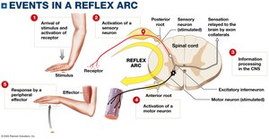
Neural Reflex: Rapid, automatic response to a specific stimulus; helps maintain homeostasis.
Reflex Arc: The pathway followed by nerve impulses to produce a reflex, typically involving sensory input, CNS integration, and motor output.
Most reflexes operate via negative feedback mechanisms.
Classification of Reflexes
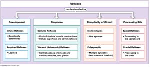
Development | Response | Complexity of Circuit | Processing Site |
|---|---|---|---|
Innate (genetically determined) | Somatic (skeletal muscle) | Monosynaptic (one synapse) | Spinal (spinal cord) |
Acquired (learned) | Visceral (smooth/cardiac muscle, glands) | Polysynaptic (multiple synapses) | Cranial (brain) |
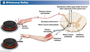
Withdrawal Reflexes
Move a body part away from a painful or threatening stimulus (e.g., touching something hot).
Involve excitatory and inhibitory interneurons to coordinate flexor and extensor muscles.
Summary Table: Major Concepts
Concept | Key Points |
|---|---|
Spinal Cord | 31 segments, bilateral symmetry, central canal, protected by meninges |
Spinal Nerves | Mixed nerves, anterior and posterior roots, dermatomes, plexuses |
Gray/White Matter | Gray: integration, nuclei, horns; White: tracts, columns, transmission |
Neuronal Pools | Divergence, convergence, serial/parallel processing, reverberation |
Reflexes | Reflex arc, classification by development, response, circuit, site |
