Back
BackThe Spinal Cord, Spinal Nerves, and the Autonomic Nervous System
Study Guide - Smart Notes
 Tailored notes based on your materials, expanded with key definitions, examples, and context.
The Spinal Cord: Structure and Organization
Gross Anatomy of the Spinal Cord
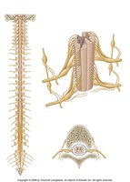
The spinal cord is a cylindrical structure that extends from the foramen magnum at the base of the skull to the level of the first or second lumbar vertebra. It serves as the main pathway for information connecting the brain and peripheral nervous system.
Conus medullaris: The tapered, lower end of the spinal cord.
Cauda equina: A bundle of spinal nerves and nerve roots extending from the conus medullaris, resembling a horse's tail.
Filum terminale: A fibrous extension of the pia mater that anchors the spinal cord to the coccyx.
Sectional Organization of the Spinal Cord
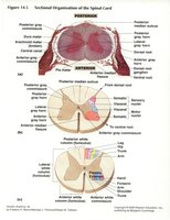
The spinal cord is organized into gray and white matter, each with distinct functions. The gray matter is centrally located and shaped like a butterfly, while the white matter surrounds it and contains ascending and descending tracts.
Posterior (dorsal) gray horn: Contains interneurons that receive somatic and visceral sensory input.
Anterior (ventral) gray horn: Contains motor neurons that innervate skeletal muscles.
Lateral gray horn: Present in thoracic and upper lumbar segments; contains autonomic (visceral motor) neurons.
White matter: Organized into columns (funiculi) containing sensory (ascending) and motor (descending) tracts.
Spinal Cord Tracts
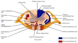
Spinal cord tracts are bundles of axons that carry specific types of information. They are classified as ascending (sensory) or descending (motor) tracts.
Ascending tracts: Carry sensory information to the brain (e.g., proprioception, pain, temperature, touch, and pressure).
Descending tracts: Transmit motor commands from the brain to the body.
Tract | Function |
|---|---|
Spinocerebellar | Proprioception to cerebellum |
Spinothalamic | Pain & temperature to thalamus |
Fasciculus gracilis/cuneatus | Touch & pressure (lower/upper body) |
Spinal Nerves and Plexuses
Spinal Nerve Structure
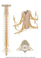
Each spinal nerve is formed by the union of an anterior (ventral) root and a posterior (dorsal) root. The roots contain motor and sensory fibers, respectively.
Anterior rootlets/root: Carry motor (efferent) fibers from the spinal cord to muscles and glands.
Posterior rootlets/root: Carry sensory (afferent) fibers from receptors to the spinal cord.
Dorsal root ganglion: Contains cell bodies of sensory neurons.
Rami: After exiting the vertebral column, each spinal nerve splits into anterior and posterior rami, which innervate different regions of the body.
Nerve Plexuses
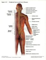
A nerve plexus is a complex network of intersecting nerves. Major plexuses include the cervical, brachial, lumbar, and sacral plexuses, which innervate the limbs and body wall.
Cervical plexus: Innervates the neck and diaphragm.
Brachial plexus: Innervates the upper limb.
Lumbar and sacral plexuses: Innervate the lower limb.
The Autonomic Nervous System (ANS)
Overview of the ANS
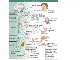
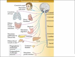
The autonomic nervous system (ANS) regulates involuntary physiological functions, including heart rate, digestion, respiratory rate, and glandular activity. It is divided into the sympathetic and parasympathetic divisions, which have opposing effects on target organs.
Sympathetic division: Prepares the body for 'fight or flight' responses; increases heart rate, dilates pupils, inhibits digestion.
Parasympathetic division: Promotes 'rest and digest' activities; slows heart rate, stimulates digestion, constricts pupils.
Anatomical differences: Sympathetic fibers originate from thoracic and lumbar regions; parasympathetic fibers arise from cranial and sacral regions.
Comparison of Sympathetic and Parasympathetic Divisions
Feature | Sympathetic | Parasympathetic |
|---|---|---|
Origin | Thoracic & lumbar spinal cord | Cranial & sacral spinal cord |
Main function | Fight or flight | Rest and digest |
Neurotransmitter (postganglionic) | Norepinephrine | Acetylcholine |
Effect on heart | Increases rate | Decreases rate |
Effect on digestion | Inhibits | Stimulates |
Clinical Application: Lumbar Puncture
Lumbar Puncture Procedure
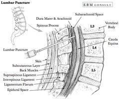
A lumbar puncture (spinal tap) is a medical procedure used to collect cerebrospinal fluid (CSF) for diagnostic testing. The needle is inserted between the L3/L4 or L4/L5 vertebrae, below the end of the spinal cord, to avoid injury to nervous tissue.
Structures traversed: Skin, subcutaneous tissue, ligaments, dura mater, and arachnoid mater to reach the subarachnoid space.
Clinical importance: Used to diagnose infections (e.g., meningitis), hemorrhage, and other neurological conditions.