 Back
BackThe Spinal Cord: Structure, Function, and Clinical Significance
Study Guide - Smart Notes
 Tailored notes based on your materials, expanded with key definitions, examples, and context.
The Spinal Cord
Overview and Functions
The spinal cord is a crucial component of the central nervous system, serving as a conduit for information between the brain and the rest of the body. It is responsible for both sensory and motor communication and acts as a major reflex center.
Two-way communication: The spinal cord transmits signals to and from the brain and body.
Reflex center: Many reflexes are initiated and completed within the spinal cord without direct involvement of the brain.
Conduction: Bundles of axons conduct sensory signals upward to the brain and motor signals downward to muscles and glands.
Gross Anatomy and Protection
The spinal cord is protected by the vertebral column, meninges, and cerebrospinal fluid (CSF). It begins at the foramen magnum and ends at the level of the first or second lumbar vertebra (L1 or L2).
Bone: The vertebral column encases the spinal cord.
Meninges: Three protective membranes surround the cord: dura mater, arachnoid mater, and pia mater.
CSF: Cerebrospinal fluid fills the subarachnoid space, providing cushioning.
Epidural space: Contains fat and veins, located between the vertebrae and dura mater.
Site of lumbar puncture: Dural and arachnoid membranes extend to the sacrum, allowing safe access for CSF sampling below the end of the cord.
Spinal Meninges
The meninges are three protective membranes that cover the brain and spinal cord, preventing abrasion and trauma.
Dura mater: Outermost, tough, collagenous membrane against bone.
Arachnoid mater: Middle layer, web-like appearance.
Pia mater: Innermost, thin membrane directly touching nervous tissue.
Too thin to see with naked eye.
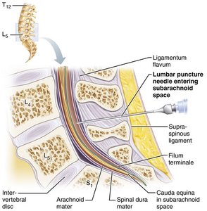
Gross Anatomy: Key Structures
The spinal cord terminates in the conus medullaris (cone shaped structure), with the filum terminale anchoring it to the coccyx.
Denticulate ligaments secure the cord laterally. Cervical and lumbar enlargements are regions where nerves serving the limbs arise.
Conus medullaris: Cone-shaped terminal end of the spinal cord.
Filum terminale: Fibrous extension covered with pia mater, anchors the cord.
Denticulate ligaments: Extensions of pia mater securing the cord to dura mater.
Cervical and lumbar enlargements: Areas where nerves for upper and lower limbs originate.
Spinal Nerves and Segmentation
Spinal nerves are part of the peripheral nervous system (PNS) and attach to the spinal cord by 31 paired roots. Each segment of the cord is designated by the paired nerves arising from it. The cauda equina is a collection of nerve roots at the inferior end of the vertebral canal.
31 pairs of spinal nerves: Attach to the cord by dorsal and ventral roots.
Cervical and lumbosacral enlargements: Nerves serving limbs emerge here.
Cauda equina: Bundle of nerve roots below the conus medullaris.
Spinal Cord Cross-Sectional Anatomy
The spinal cord is divided into right and left halves by two grooves: the ventral (anterior) median fissure and the dorsal (posterior) median sulcus. Gray matter is located centrally, surrounded by white matter. The central canal runs the length of the cord and is filled with CSF.
Gray matter: Core of the cord, shaped like a butterfly or "H".
White matter: Surrounds the gray matter, contains myelinated and unmyelinated fibers.
Central canal: Contains CSF.
Gray Matter and Spinal Roots
Gray matter is organized into dorsal, ventral, and lateral horns. Dorsal horns contain interneurons receiving sensory input; ventral horns contain motor neurons; lateral horns (in thoracic and superior lumbar regions) contain sympathetic neurons. The gray commissure connects the two sides and encloses the central canal.
Dorsal horns: Receive somatic and visceral sensory input.
Ventral horns: Contain somatic motor neurons.
Lateral horns: Contain sympathetic neurons (only in certain regions).
Gray commissure: Bridge connecting gray matter on both sides.
Ventral roots: Motor neuron axons exiting the cord.
Dorsal roots: Sensory input entering the cord.
Dorsal root ganglia: Cell bodies of sensory neurons.
Spinal nerves: Formed by fusion of dorsal and ventral roots.
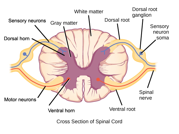
Functional Groups in Gray Matter
Gray matter is divided into four functional groups:
Somatic Sensory (SS): Receives input from somatic sensory neurons.
Visceral Sensory (VS): Receives input from visceral sensory neurons.
Visceral (autonomic) Motor (VM): Contains visceral motor neurons.
Somatic Motor (SM): Contains somatic motor neurons.
Spinal Cord White Matter
White matter consists of myelinated and unmyelinated fibers that allow communication between different parts of the spinal cord and between the cord and brain. Fibers run in three directions:
Ascending: Carry sensory information to higher levels of the cord or brain.
Descending: Carry motor signals from the brain down the cord.
Transverse: Tracts cross over from one side of the body to the other.
This crossover explains why strokes on one side of the brain affect the opposite side of the body.
Spinal Cord Trauma and Disorders
Injury to the spinal cord or its roots can lead to significant functional losses. The effects depend on the location and severity of the injury.
Paresthesia: Loss of sensory function due to damage to dorsal roots or sensory tracts.
Paralysis: Loss of motor function due to damage to ventral roots or ventral horn cells.
Flaccid paralysis: Severe damage; no voluntary or involuntary control; muscles atrophy.
Spastic paralysis: Damage to upper motor neurons; muscles stimulated by reflex activity; no voluntary control; muscles may shorten permanently.
Transection: Complete cut of the cord results in total motor and sensory loss below the injury.
Paraplegia: Transection between T1 and L1; loss of function in lower limbs.
Quadriplegia: Transection in cervical region; loss of function in all limbs.
Spinal shock: Temporary loss of function below the lesion.
Amyotrophic lateral sclerosis (ALS): Progressive destruction of motor neurons; loss of ability to speak, swallow, and breathe; typically fatal within five years.
Clinical significance: Spinal cord injuries are serious because they disrupt communication between the brain and body, leading to paralysis and loss of function.
Type of Injury | Effect |
|---|---|
Paresthesia | Sensory function loss |
Flaccid paralysis | No voluntary/involuntary control; muscle atrophy |
Spastic paralysis | No voluntary control; reflex activity remains |
Transection (T1-L1) | Paraplegia (lower limbs affected) |
Transection (cervical) | Quadriplegia (all limbs affected) |
ALS | Progressive motor neuron loss; fatal |
Example: A spinal cord injury at the cervical level can result in quadriplegia, affecting all four limbs and potentially respiratory function.
Additional info: The spinal cord is essential for both voluntary and involuntary actions, and its damage can have profound effects on quality of life and survival.
