 Back
The Spinal Cord: Structure, Function, and Pathways (CNS Part B)
Study Guide - Smart Notes
 Tailored notes based on your materials, expanded with key definitions, examples, and context.
The Spinal Cord: Structure, Function, and Homeostasis
Overview and Functions
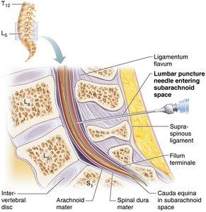
The spinal cord is a critical component of the central nervous system (CNS), providing two-way communication between the brain and the body. It serves as the major reflex center, where reflexes are initiated and completed. The spinal cord is protected by the vertebral column and extends from the foramen magnum to the level of the L1 or L2 vertebra, terminating in the conus medullaris. The filum terminale anchors the cord to the coccyx, and denticulate ligaments secure it laterally. Enlargements in the cervical and lumbar regions accommodate nerves supplying the limbs, while the cauda equina is a bundle of nerve roots at the inferior end.
Protection of the Spinal Cord
Bone: The vertebral column encases and protects the spinal cord.
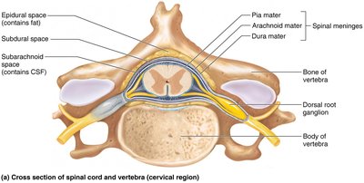
Meninges: Three connective tissue membranes (dura mater, arachnoid mater, pia mater) surround the cord.
Cerebrospinal Fluid (CSF): Fills the subarachnoid space, providing cushioning and buoyancy.z
Epidural Space: Contains fat and veins between the vertebrae and dura mater.
Clinical Note: The dural and arachnoid membranes extend to the sacrum, making the lower lumbar region (below L2) a safe site for lumbar puncture (spinal tap).
Spinal Cord Cross-Sectional Anatomy
General Structure
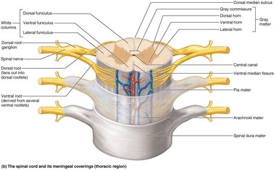
The spinal cord is partially divided into right and left halves by the ventral (anterior) median fissure and the dorsal (posterior) median sulcus. The central canal, filled with CSF, runs the length of the cord. Gray matter forms the core, while white matter surrounds it.
Gray Matter and Spinal Roots
Shape: The gray matter resembles a butterfly or the letter "H" in cross-section.
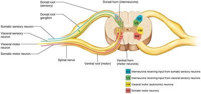
Regions: Each side contains dorsal horns (interneurons for sensory input), ventral horns (motor neurons), and lateral horns (sympathetic neurons in thoracic/superior lumbar regions).
Gray Commissure: Connects the two sides of gray matter and encloses the central canal.
Roots: Ventral roots carry motor axons out; dorsal roots bring sensory input in. Dorsal root ganglia house sensory neuron cell bodies. Spinal nerves are formed by the fusion of dorsal and ventral roots.
Functional Groups: Gray matter is divided into four regions based on function: Somatic sensory (SS), Visceral sensory (VS), Visceral motor (VM), and Somatic motor (SM).
White Matter
Composition: Myelinated and unmyelinated fibers allow communication within the spinal cord and between the cord and brain.
Directions: Fibers run ascending (sensory), descending (motor), and transversely (commissural).
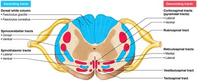
Columns (Funiculi): White matter is organized into dorsal (posterior), lateral, and ventral (anterior) columns. Each column contains tracts of axons with similar functions and destinations.
Neuronal Pathways in the Spinal Cord
General Principles
Decussation: Most pathways cross from one side of the CNS to the other.
Relay: Pathways typically consist of two or three neurons in sequence.
Somatotopy: Spatial relationships in the CNS reflect those in the body.
Symmetry: Pathways are paired on both sides of the spinal cord.
Ascending (Sensory) Pathways
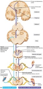
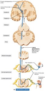
Ascending pathways conduct sensory information upward through a chain of three neurons:
First-order neuron: Conducts impulses from receptors to the spinal cord or medulla.
Second-order neuron: Interneuron with cell body in the dorsal horn or medullary nuclei; axons extend to the thalamus or cerebellum.
Third-order neuron: Interneuron with cell body in the thalamus; axons project to the somatosensory cortex (not present in cerebellar pathways).
Main Ascending Pathways
Dorsal column–medial lemniscal pathways: Transmit discriminative touch and vibration to the somatosensory cortex via the thalamus. Composed of the fasciculus cuneatus, fasciculus gracilis, and medial lemniscus.
Spinothalamic pathways: Lateral and ventral tracts transmit pain, temperature, coarse touch, and pressure.
Spinocerebellar tracts: Ventral and dorsal tracts convey muscle/tendon stretch information to the cerebellum for coordination of muscle activity.
Descending (Motor) Pathways and Tracts
Descending pathways deliver efferent impulses from the brain to the spinal cord and are divided into two groups:
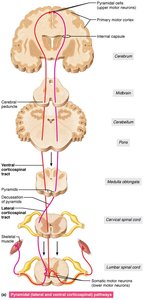
Direct (pyramidal) pathways: Originate from pyramidal cells in the primary motor cortex and descend via corticospinal tracts. These regulate fast, fine (skilled) movements. Axons descend without synapsing until reaching the spinal cord, where they synapse with interneurons or ventral horn motor neurons.
Indirect (extrapyramidal) pathways: All other motor pathways, including those from brainstem motor nuclei. These are complex, multisynaptic, and regulate axial muscles (posture/balance), coarse limb movements, and head/neck/eye movements in response to visual stimuli.
Major Indirect Pathways
Reticulospinal and vestibulospinal tracts: Maintain balance by adjusting postural muscle tone.
Rubrospinal tracts: Control flexor muscles.
Tectospinal tracts: Mediate head movements in response to visual stimuli.
Summary Table: Major Spinal Cord Tracts
Tract | Type | Main Function |
|---|---|---|
Dorsal column–medial lemniscal | Ascending (sensory) | Discriminative touch, vibration |
Spinothalamic | Ascending (sensory) | Pain, temperature, coarse touch, pressure |
Spinocerebellar | Ascending (sensory) | Muscle/tendon stretch (proprioception) |
Corticospinal (pyramidal) | Descending (motor) | Fine, skilled voluntary movement |
Reticulospinal, vestibulospinal | Descending (motor, indirect) | Posture, balance |
Rubrospinal | Descending (motor, indirect) | Flexor muscle control |
Tectospinal | Descending (motor, indirect) | Head/neck movement in response to visual stimuli |
Key Terms and Concepts
Conus medullaris: Tapered, terminal end of the spinal cord.
Cauda equina: Bundle of nerve roots at the lower end of the spinal cord.
Filum terminale: Fibrous extension anchoring the spinal cord to the coccyx.
Meninges: Dura mater, arachnoid mater, pia mater—protective coverings of the CNS.
Decussation: Crossing of nerve fibers from one side to the other.
Somatotopy: Point-for-point correspondence between body regions and CNS regions.
References: Marieb, E. N., & Hoehn, K. (2023). Human Anatomy and Physiology (12th ed.). Pearson.
