 Back
BackThe Spinal Cord: Structure, Meninges, and Functional Pathways
Study Guide - Smart Notes
 Tailored notes based on your materials, expanded with key definitions, examples, and context.
The Meninges of the Spinal Cord
Protective Layers and Spaces
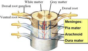
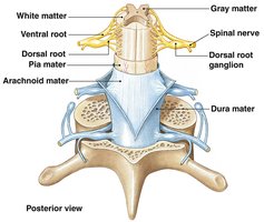
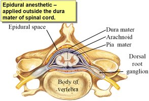
The spinal cord is protected by three connective tissue membranes known as the meninges, which are continuous with those of the brain. These layers provide structural support, protection, and a conduit for blood vessels.
Dura mater: The tough, outermost layer.
Arachnoid mater: The middle, web-like layer.
Pia mater: The delicate, innermost layer that adheres closely to the spinal cord.
Epidural space: The area outside the dura mater, containing fat and blood vessels.
Subdural space: A potential space between the dura and arachnoid mater.
Subarachnoid space: The space between the arachnoid and pia mater, filled with cerebrospinal fluid (CSF).
Meningitis is the inflammation of the meninges, often caused by infection.
Spinal Cord Anatomy
Gross Structure and Regions
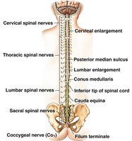
The spinal cord extends from the brainstem to the level of the first or second lumbar vertebra (L1–L2). It serves as a major pathway for information traveling between the brain and the peripheral nervous system (PNS).
31 pairs of spinal nerves emerge from the spinal cord, each serving a specific region of the body.
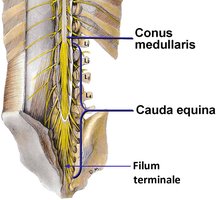
Conus medullaris: The tapered, cone-shaped end of the spinal cord, typically at L1.
Cauda equina: A bundle of spinal nerves extending below the conus medullaris, resembling a horse's tail.
Filum terminale: A fibrous extension of the pia mater that anchors the spinal cord to the coccyx.
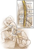
Lumbar Puncture and Epidural Anesthesia
Clinical Procedures Involving the Spinal Cord
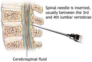
Lumbar puncture (spinal tap) is a procedure used to withdraw cerebrospinal fluid (CSF) for diagnostic purposes or to administer medications. The needle is typically inserted between the L3 and L4 vertebrae, below the conus medullaris, to avoid damaging the spinal cord.
CSF analysis can help diagnose infections such as meningitis.
Epidural injection: Medication is administered into the epidural space for pain relief, commonly during childbirth.
Spinal anesthesia: Medication is injected into the subarachnoid space, causing loss of sensation.
Spinal analgesia: Medication for pain relief, often used in combination with anesthetics.
Functions of the Spinal Cord
Major Roles in the Nervous System
The spinal cord is essential for transmitting sensory and motor information and for mediating reflexes.
Receives sensory input from peripheral tissues via sensory neurons.
Transmits sensory information to the brain through ascending tracts.
Relays motor commands from the brain via descending tracts to peripheral effectors.
Controls basic reflexes independent of the brain.
Internal Anatomy of the Spinal Cord
Gray and White Matter Organization
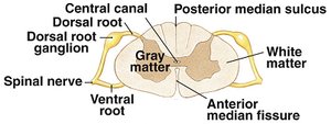
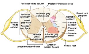
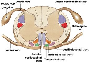
The spinal cord consists of central gray matter (containing neuron cell bodies) and surrounding white matter (containing myelinated axons).
Gray matter: Organized into horns and nuclei for processing sensory and motor information.
White matter: Organized into columns containing ascending (sensory) and descending (motor) tracts.
Organization of Gray Matter
Gray matter is divided into regions (horns) and nuclei, each associated with specific functions:
Somatic sensory nuclei: Relay information from skin, muscles, and joints.
Visceral sensory nuclei: Relay information from internal organs.
Visceral motor nuclei: Send commands to smooth muscle, cardiac muscle, and glands.
Somatic motor nuclei: Send commands to skeletal muscles.
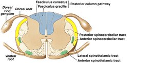
Ascending (Sensory) Tracts of the Spinal Cord
Major Sensory Pathways
Ascending tracts in the white matter carry sensory information from the body to the brain. The three major pathways are:
Posterior column (medial lemniscus) pathway: Fine touch and conscious proprioception.
Spinothalamic tracts: Pain, temperature, crude touch, and pressure.
Spinocerebellar tracts: Subconscious proprioception (muscle sense).
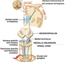
Posterior Column Pathway
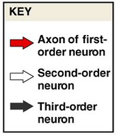
This pathway transmits fine touch and conscious proprioception to the brain. It involves a sequence of three neurons: first-order, second-order, and third-order neurons.
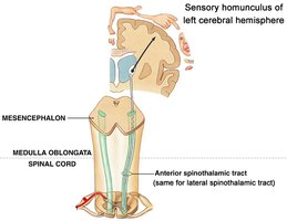
Spinothalamic Tract
This tract carries information about crude touch, pressure, pain, and temperature. It also involves a three-neuron chain.

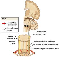
Spinocerebellar Tract
This tract conveys subconscious proprioceptive information from muscles and joints to the cerebellum, aiding in coordination and balance.
Descending (Motor) Tracts of the Spinal Cord
Major Motor Pathways
Descending tracts carry motor commands from the brain to the body. The two main types are:
Corticospinal (pyramidal) tracts: Responsible for voluntary muscle control.
Extrapyramidal tracts: Involved in balance, muscle tone, and reflexes.
Somatic Nervous System: Conscious vs. Subconscious Motor Control
The somatic nervous system controls skeletal muscles through both conscious and subconscious pathways:
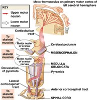
Conscious motor control: Originates in the primary motor cortex and travels via corticospinal tracts.
Subconscious motor control: Originates in the brainstem and travels via extrapyramidal tracts, adjusting balance, muscle tone, and posture.
Corticospinal (Pyramidal) Tracts
These tracts carry voluntary motor commands from the cortex to skeletal muscles, crossing over (decussating) in the medulla oblongata.
Additional info: The spinal cord is a critical structure for both sensory and motor integration, and its organization into specific tracts and nuclei allows for precise control and communication between the brain and body. Damage to specific tracts or regions can result in characteristic patterns of sensory or motor loss, which is important in clinical diagnosis.
