Back
BackThe Spinal Nerves: Structure, Plexuses, and Functions
Study Guide - Smart Notes
 Tailored notes based on your materials, expanded with key definitions, examples, and context.
The Spinal Nerves
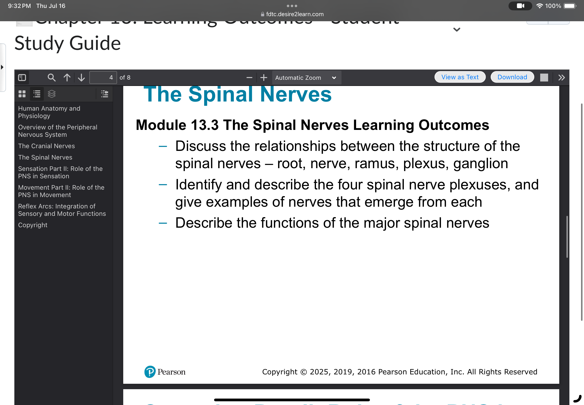
Structure of the Spinal Nerves
The spinal nerves are essential components of the peripheral nervous system (PNS), connecting the central nervous system (CNS) to limbs and organs. Their structure is organized into several distinct parts, each with specific functions.
Root: The spinal nerve roots are the initial segments that emerge from the spinal cord. There are two types: dorsal (posterior) root (sensory) and ventral (anterior) root (motor).
Nerve: The spinal nerve is formed by the union of the dorsal and ventral roots. It is a mixed nerve, carrying both sensory and motor fibers.
Ramus: After exiting the vertebral column, each spinal nerve splits into branches called rami. The dorsal ramus serves the muscles and skin of the back, while the ventral ramus supplies the limbs and anterior trunk.
Plexus: The ventral rami of several spinal nerves combine to form nerve plexuses, which are networks that redistribute fibers to form peripheral nerves.
Ganglion: A ganglion is a collection of neuron cell bodies located outside the CNS. The dorsal root ganglion contains sensory neuron cell bodies.
The Four Spinal Nerve Plexuses
Spinal nerve plexuses are complex networks formed by the ventral rami of spinal nerves. They allow for the redistribution of nerve fibers, ensuring that each peripheral nerve contains fibers from multiple spinal levels.
Cervical Plexus: Formed by C1-C5. Example: Phrenic nerve (controls the diaphragm).
Brachial Plexus: Formed by C5-T1. Example: Radial nerve (innervates the posterior arm and forearm).
Lumbar Plexus: Formed by L1-L4. Example: Femoral nerve (innervates anterior thigh muscles).
Sacral Plexus: Formed by L4-S4. Example: Sciatic nerve (largest nerve, innervates lower limb).
Functions of Major Spinal Nerves
Major spinal nerves are responsible for transmitting sensory and motor information between the CNS and the rest of the body. Their functions are determined by the regions they serve.
Phrenic nerve: Controls the diaphragm, essential for breathing.
Radial nerve: Enables extension of the wrist and fingers.
Femoral nerve: Facilitates hip flexion and knee extension.
Sciatic nerve: Provides motor and sensory functions to the posterior thigh, leg, and foot.
Example:
The sciatic nerve is clinically significant because injury can lead to weakness or loss of sensation in the lower limb.
Additional info:
Spinal nerves are classified as mixed nerves because they contain both afferent (sensory) and efferent (motor) fibers. The organization into plexuses allows for redundancy and protection against injury, as damage to a single spinal segment does not necessarily result in complete loss of function in a limb.