 Back
BackThe Synapse: Structure, Function, and Integration in the Nervous System
Study Guide - Smart Notes
 Tailored notes based on your materials, expanded with key definitions, examples, and context.
The Synapse
Definition and Functional Overview
The synapse is a specialized junction that facilitates the transfer of information from one neuron to another or from a neuron to an effector cell. Synapses are essential for neural communication, allowing the nervous system to process and transmit signals efficiently.
Presynaptic neuron: The neuron sending the signal.
Postsynaptic neuron: The neuron receiving the signal.
Synapses can also occur between a neuron and a muscle or gland cell (effector cell).
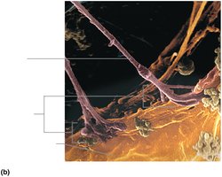
Types of Synaptic Connections
Neurons can connect in several ways, depending on the location of the synapse:
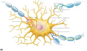
Axodendritic: Between axon terminals of one neuron and dendrites of another.
Axosomatic: Between axon terminals and the soma (cell body) of another neuron.
Axoaxonal, dendrodendritic, somatodendritic: Less common, involving axon-to-axon, dendrite-to-dendrite, or dendrite-to-soma connections.
Types of Synapses
Chemical Synapses
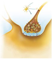
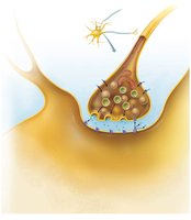
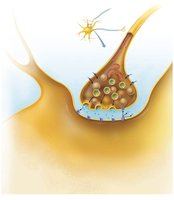
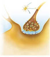
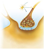
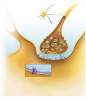
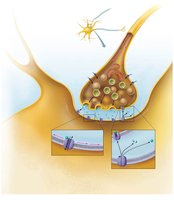
Chemical synapses are the most common type in the nervous system. They use neurotransmitters to transmit signals across a fluid-filled synaptic cleft, ensuring unidirectional communication.
Axon terminal: Contains synaptic vesicles filled with neurotransmitter.
Synaptic cleft: Small gap separating the presynaptic and postsynaptic membranes.
Receptor region: Located on the postsynaptic membrane, binds neurotransmitter.
Electrical Synapses
Electrical synapses are less common and involve direct cytoplasmic connections via gap junctions. They allow rapid, bidirectional transmission of electrical signals and are found in certain brain regions and embryonic tissue.
Transmission Across a Chemical Synapse
Steps of Synaptic Transmission
The process of signal transmission at a chemical synapse involves several key steps:
Action potential arrives at the axon terminal.
Voltage-gated Ca2+ channels open; Ca2+ enters the terminal.
Ca2+ influx triggers exocytosis of neurotransmitter vesicles.
Neurotransmitter diffuses across the synaptic cleft and binds to receptors on the postsynaptic membrane.
Binding opens ligand-gated ion channels, generating a graded potential.
Neurotransmitter effects are terminated by reuptake, enzymatic degradation, or diffusion away from the cleft.
Neurotransmitters
Definition and Classification
Neurotransmitters are chemical messengers that neurons use to communicate at chemical synapses. Over 50 neurotransmitters have been identified, and they are classified by chemical structure and function.
Acetylcholine (ACh): Used at neuromuscular junctions, degraded by acetylcholinesterase.
Biogenic amines: Includes catecholamines (dopamine, norepinephrine, epinephrine) and indolamines (serotonin, histamine).
Amino acids: Glutamate, aspartate, glycine, GABA.
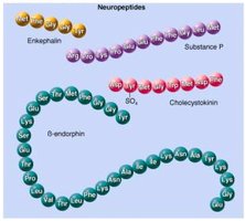
Peptides (neuropeptides): Substance P, endorphins, somatostatin, cholecystokinin.
Purines: ATP, adenosine.
Gases and lipids: Nitric oxide (NO), carbon monoxide (CO), hydrogen sulfide (H2S), endocannabinoids.
Neurotransmitter Actions
Excitatory (depolarizing): Promote action potential generation (e.g., glutamate).
Inhibitory (hyperpolarizing): Suppress action potential generation (e.g., GABA, glycine).
Direct action: Bind to and open ion channels (e.g., ACh, amino acids).
Indirect action: Act through second messengers and G protein-coupled receptors (e.g., biogenic amines, neuropeptides).
Postsynaptic Potentials
Excitatory and Inhibitory Postsynaptic Potentials (EPSPs and IPSPs)
Neurotransmitter binding to postsynaptic receptors generates graded potentials:
EPSP (Excitatory Postsynaptic Potential): Local depolarization, brings neuron closer to threshold for firing an action potential.
IPSP (Inhibitory Postsynaptic Potential): Local hyperpolarization, moves neuron further from threshold, decreasing likelihood of firing.
Summation and Integration
Neurons integrate thousands of synaptic inputs through summation:
Temporal summation: Rapid, repeated signals from one presynaptic neuron.
Spatial summation: Simultaneous signals from multiple presynaptic neurons.
EPSPs and IPSPs can add together, and only if threshold is reached will an action potential be generated.
Neural Integration and Processing
Neuronal Pools and Circuits
Neurons are organized into neuronal pools and circuits that process information:
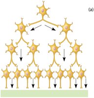
Diverging circuit: One input, many outputs (amplification).
Converging circuit: Many inputs, one output (concentration).
Reverberating circuit: Signal travels in a loop, producing rhythmic activity.
Parallel after-discharge circuit: Signal stimulates neurons in parallel, causing a burst of output.
Serial and Parallel Processing
Serial processing: Input travels along one pathway to a specific destination (e.g., reflex arc).
Parallel processing: Input travels along several pathways, allowing complex responses and higher-level processing.
Developmental Aspects of Neurons
Neural Development and Synapse Formation
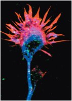
The nervous system originates from the neural tube and neural crest. Neurons form connections through axonal growth cones, guided by adhesion proteins and neurotrophic factors. Synaptic connections are refined during development, with unused neurons undergoing apoptosis.
Additional info: During childhood and adolescence, synaptic pruning and reinforcement shape neural circuits, and some neuronal populations (e.g., olfactory neurons, hippocampus) retain the ability to divide throughout life.
