 Back
BackThe Thoracic Cage and Vertebral Column: Structure, Function, and Disorders
Study Guide - Smart Notes
 Tailored notes based on your materials, expanded with key definitions, examples, and context.
The Thoracic Cage
Overview of the Thoracic Cage
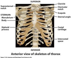
The thoracic cage, also known as the rib cage, is a bony structure that protects vital organs in the thoracic cavity, such as the heart and lungs. It is composed of the sternum, ribs, and thoracic vertebrae, and provides attachment points for muscles involved in respiration and upper limb movement.
Sternum (Breastbone): A flat bone located at the anterior midline of the thorax, consisting of three parts: the manubrium (superior), body (middle and largest), and xiphoid process (smallest and most inferior).
Ribs: Twelve pairs of curved bones that form the lateral aspects of the thoracic cage. They connect posteriorly to the thoracic vertebrae and anteriorly to the sternum via costal cartilages.
Costal Cartilage: Hyaline cartilage that connects the ribs to the sternum, allowing flexibility and expansion during breathing.
Classification of Ribs
True Ribs (Vertebrosternal, Pairs 1-7): Attach directly to the sternum via their own costal cartilages.
False Ribs (Vertebrochondral, Pairs 8-10): Attach to the sternum indirectly by joining the cartilage of the rib above.
Floating Ribs (Vertebral, Pairs 11-12): Do not attach to the sternum at all; only attached posteriorly to the thoracic vertebrae.
Example: Ribs 1-7 are true ribs, ribs 8-10 are false ribs, and ribs 11-12 are floating ribs.
Structure of a Typical Rib
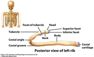
Each rib has several anatomical features that facilitate articulation with the vertebrae and sternum.
Head: Articulates with the bodies of thoracic vertebrae.
Neck: Connects the head to the body of the rib.
Tubercle: Articulates with the transverse process of the vertebra.
Body (Shaft): Main part of the rib, curved and thin.
Costal Groove: Houses nerves and blood vessels.
Articulation of Ribs with Vertebrae and Sternum
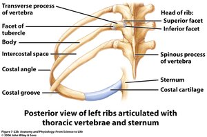
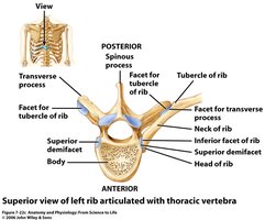
Ribs articulate posteriorly with thoracic vertebrae and anteriorly with the sternum (directly or indirectly).
The head of the rib articulates with the vertebral body.
The tubercle of the rib articulates with the transverse process of the vertebra.
Costal cartilage connects the anterior end of most ribs to the sternum.
The Vertebral Column
Regions of the Vertebral Column
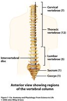
The vertebral column, or spine, is a flexible, segmented structure that supports the body and protects the spinal cord. It is divided into five regions:
Cervical (C1-C7): Seven vertebrae in the neck region.
Thoracic (T1-T12): Twelve vertebrae posterior to the thoracic cavity.
Lumbar (L1-L5): Five vertebrae supporting the lower back.
Sacrum: Five fused sacral vertebrae forming the posterior wall of the pelvis.
Coccyx: Three to five fused coccygeal vertebrae forming the tailbone.
Curvatures of the Vertebral Column
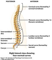
The vertebral column has four natural curves that develop over time, contributing to its strength, flexibility, and ability to absorb shock.
Primary Curves: Thoracic and sacral curves, present from fetal development.
Secondary Curves: Cervical and lumbar curves, develop after birth as the infant begins to hold up the head and walk.
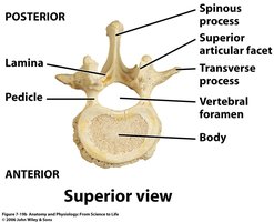
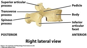
Structure of a Typical Vertebra
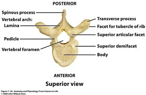
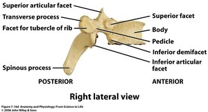
Each vertebra consists of several key structures that provide support and allow for articulation with adjacent vertebrae and ribs.
Body: The weight-bearing portion.
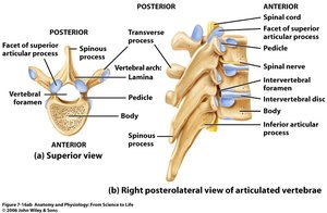
Vertebral Arch: Formed by the pedicle and lamina, enclosing the vertebral foramen (spinal canal).
Processes: Transverse, spinous, and articular processes serve as attachment points for muscles and ligaments.
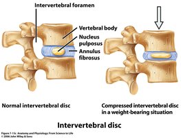
Intervertebral Foramen: Openings between vertebrae for the passage of spinal nerves.
Intervertebral Discs
Intervertebral discs are fibrocartilaginous pads located between adjacent vertebral bodies. They act as shock absorbers and allow for flexibility in the spine.
Annulus Fibrosus: Outer ring of fibrocartilage.
Nucleus Pulposus: Gelatinous core that provides cushioning.
Disorders of the Vertebral Column
Kyphosis, Lordosis, and Scoliosis
Abnormal curvatures of the spine can result in various clinical conditions:
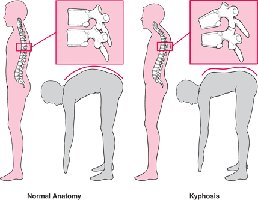
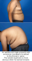
Kyphosis: Excessive posterior curvature of the thoracic region (hunchback). Causes include osteoporosis and repetitive motion.
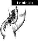
Lordosis: Excessive anterior curvature of the lumbar region (swayback). Causes include pregnancy, obesity, and weak abdominal muscles.
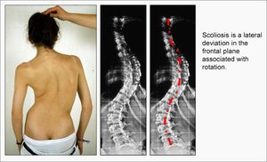
Scoliosis: Lateral curvature of the spine, which may be due to habitual, structural, or idiopathic causes.
Spina Bifida
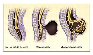
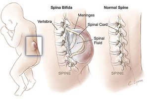
Spina bifida is a congenital defect where the lamina of the vertebra fails to fuse properly, resulting in exposure of the spinal cord and/or meninges.
Spina Bifida Occulta: Mildest form, usually affects the fifth lumbar vertebra and causes no symptoms.
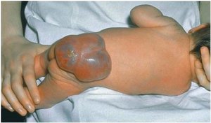
Meningocele: Meninges protrude through the defect.
Myelomeningocele: Both the spinal cord and meninges protrude, often resulting in neurological deficits.
Regional Specializations of the Vertebral Column
Cervical Vertebrae
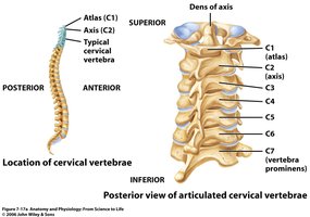
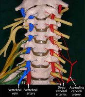
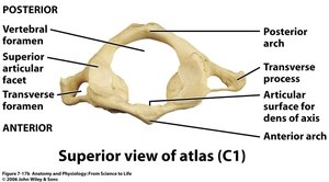
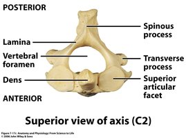
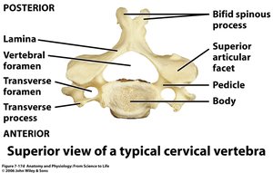
The cervical region consists of seven vertebrae (C1-C7). Unique features include the transverse foramen (for vertebral arteries) and bifid spinous processes. The atlas (C1) and axis (C2) are specialized for head movement.
Atlas (C1): Supports the skull; allows nodding motion.
Axis (C2): Contains the dens (odontoid process) for rotation of the head.
Thoracic Vertebrae
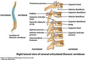
The thoracic region contains twelve vertebrae (T1-T12), each articulating with a pair of ribs. They have long, downward-pointing spinous processes and facets for rib articulation.
Lumbar Vertebrae
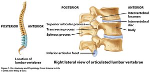
The lumbar region consists of five vertebrae (L1-L5). These vertebrae are the largest and thickest, designed to bear the weight of the upper body.
Sacrum and Coccyx
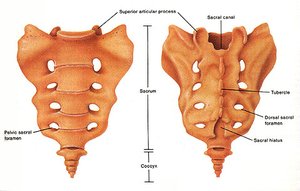
The sacrum is composed of five fused vertebrae, forming the posterior wall of the pelvis. The coccyx, or tailbone, consists of 3-5 fused vertebrae. The sacrum articulates with the ilium of the pelvis at the sacroiliac joints.
Transverse Lines: Indicate fusion points of sacral vertebrae.
Sacral Foramina: Openings for the passage of sacral nerves.
Sacral Hiatus: Inferior opening of the sacral canal.
Summary Table: Vertebral Regions and Key Features
Region | Number of Vertebrae | Key Features |
|---|---|---|
Cervical | 7 | Transverse foramen, bifid spinous process, atlas (C1), axis (C2) with dens |
Thoracic | 12 | Facets for ribs, long spinous process |
Lumbar | 5 | Large, thick bodies, weight-bearing |
Sacrum | 5 (fused) | Transverse lines, sacral foramina, sacral hiatus |
Coccyx | 3-5 (fused) | Small, tailbone |
