 Back
The Tissue Level of Organization: Epithelial and Connective Tissues
Study Guide - Smart Notes
 Tailored notes based on your materials, expanded with key definitions, examples, and context.
The Tissue Level of Organization
Introduction to Tissues
Tissues are collections of similar cells and cell products that perform specific functions in the body. The study of tissues is known as histology. There are four primary tissue types in the human body: epithelial, connective, muscle, and nervous tissues. These tissues combine to form organs, each with specialized functions.
Epithelial Tissue: Covers surfaces, lines cavities, and forms glands.
Connective Tissue: Supports, binds, and protects other tissues and organs.
Muscle Tissue: Specialized for contraction and movement.
Nervous Tissue: Conducts electrical impulses for communication.
Epithelial Tissue
Characteristics and Functions
Epithelial tissue consists of closely packed cells with minimal extracellular material. It is avascular (lacks blood vessels) and is anchored to underlying tissues by a basement membrane. Epithelial tissues serve several key functions:
Physical protection: Against abrasion, dehydration, and chemical or biological agents.
Control permeability: Regulate absorption and secretion of substances.
Provide sensation: Contain sensory nerve endings.
Produce secretions: Through glandular cells.
Structural Features
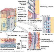
Basement membrane: Composed of the basal lamina (attaches to epithelium) and reticular lamina (deeper layer).
Cell junctions: Specialized connections between cells, including tight junctions, gap junctions, and desmosomes.
Classification of Epithelia
Epithelia are classified by the number of cell layers and the shape of the cells at the apical surface:
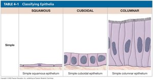
Simple: One layer of cells; all touch the basement membrane.
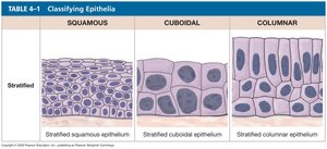
Stratified: Multiple layers; only the deepest layer touches the basement membrane.
Squamous | Cuboidal | Columnar | |
|---|---|---|---|
Simple | Simple squamous | Simple cuboidal | Simple columnar |
Stratified | Stratified squamous | Stratified cuboidal | Stratified columnar |
Types of Epithelial Tissue
Simple Squamous Epithelium
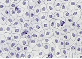
Composed of a single layer of flat, oval cells. Functions in diffusion and filtration. Found lining blood vessels (endothelium), alveoli of lungs, and body cavities (mesothelium).
Simple Cuboidal Epithelium
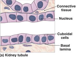
Single layer of cube-shaped cells with centrally located nuclei. Functions in secretion and absorption. Located in kidney tubules, glands, and ducts.
Simple Columnar Epithelium
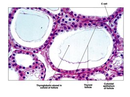
Single layer of elongated cells with nuclei near the basal lamina. May contain goblet cells (secrete mucus) and microvilli (increase surface area). Functions in absorption, secretion, and protection. Found in the digestive tract and uterine tubes.
Stratified Squamous Epithelium
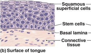
Multiple layers of flattened cells. Provides protection against abrasion. Found in the skin, mouth, throat, esophagus, vagina, and anal canal.
Stratified Cuboidal Epithelium
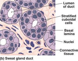
Two or more layers of cube-shaped cells. Functions in protection. Found lining ducts of sweat glands and mammary glands.
Stratified Columnar Epithelium
Several layers of cells; superficial cells are columnar. Functions in protection. Found in parts of the urethra, anus, and pharynx.
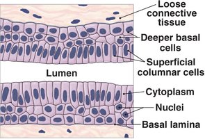
Transitional Epithelium
Multiple layers of cells with curved apical surfaces. Tolerates stretching and recoiling. Found in the urinary bladder and ureters.
Pseudostratified Ciliated Columnar Epithelium
Appears layered due to varying positions of nuclei, but all cells touch the basement membrane. Contains cilia and goblet cells. Functions in secretion and movement of mucus. Found in the respiratory tract (nasal cavity, trachea, bronchi).
Glandular Epithelium
Specialized for secretion. Glands are classified as exocrine (secrete into ducts) or endocrine (secrete hormones into blood). Modes of secretion include merocrine, apocrine, and holocrine.
Merocrine: Secretion by exocytosis (e.g., sweat glands).
Apocrine: Secretion with loss of cytoplasm (e.g., mammary glands).
Holocrine: Secretion by cell rupture (e.g., sebaceous glands).
Connective Tissue
Characteristics and Functions
Connective tissues are the most diverse and abundant tissues in the body. They have fewer cells than epithelial tissues and contain an extensive extracellular matrix composed of protein fibers and ground substance. Functions include binding, supporting, protecting, storing energy, and transporting substances.
Major fiber types: Collagen (strength), elastic (stretch), reticular (supportive networks).
Major cell types: Fibroblasts, fibrocytes, macrophages, adipocytes, mesenchymal cells, melanocytes, mast cells, lymphocytes.
Types of Connective Tissue
Loose connective tissue: Areolar, adipose, reticular.
Dense connective tissue: Regular, irregular, elastic.
Cartilage: Hyaline, elastic, fibrocartilage.
Other: Blood, lymph, bone.
Areolar Connective Tissue
Loose arrangement of fibers and cells. Supports and binds other tissues, allows for movement, and provides a reservoir for water and salts. Found under the skin and around organs.
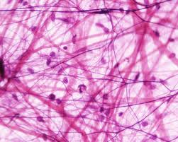
Adipose Tissue
Stores fat in vacuoles, cushions organs, and insulates the body. Found under the skin, around eyes, and in breasts.
Reticular Tissue
Forms a supportive framework for organs such as the liver, spleen, and lymph nodes.
Dense Regular Connective Tissue
Composed of tightly packed collagen fibers arranged in parallel. Provides strong attachment between structures. Found in tendons and ligaments.
Dense Irregular Connective Tissue
Collagen fibers arranged in an irregular pattern. Provides strength and support in multiple directions. Found in the dermis and organ capsules.
Elastic Tissue
Dominated by elastic fibers, allowing tissues to stretch and recoil. Found in the walls of large arteries and vertebral ligaments.
Blood and Lymph
Blood transports gases, nutrients, and wastes; contains erythrocytes, leukocytes, and platelets. Lymph returns excess tissue fluid to the bloodstream and is involved in immune responses.
Cartilage
Cartilage is a firm, flexible tissue with chondrocytes in lacunae. Types include hyaline (most common, reduces friction), elastic (flexible), and fibrocartilage (strong, resists compression).
Bone
Bone tissue is rigid due to mineralized matrix. Osteocytes reside in lacunae and communicate via canaliculi. Functions include support, protection, and blood cell production.
Membranes
Types of Membranes
Membranes are physical barriers composed of epithelial and connective tissues. Types include:
Mucous membranes: Line passageways open to the exterior (digestive, respiratory, urinary, reproductive tracts).
Serous membranes: Line closed body cavities; secrete serous fluid to reduce friction (pleura, pericardium, peritoneum).
Cutaneous membrane: The skin; protects the body surface.
Synovial membranes: Line joint cavities; produce synovial fluid for lubrication.
Summary Table: Epithelial Tissue Types
Type | Structure | Function | Location |
|---|---|---|---|
Simple Squamous | Single layer, flat cells | Diffusion, filtration | Alveoli, blood vessels |
Simple Cuboidal | Single layer, cube-shaped | Secretion, absorption | Kidney tubules, glands |
Simple Columnar | Single layer, tall cells | Absorption, secretion | Digestive tract |
Stratified Squamous | Multiple layers, flat cells | Protection | Skin, mouth, esophagus |
Stratified Cuboidal | 2+ layers, cube-shaped | Protection | Sweat glands |
Stratified Columnar | 2+ layers, tall cells | Protection | Urethra, pharynx |
Transitional | Multiple layers, variable shape | Stretching | Bladder, ureters |
Pseudostratified Ciliated Columnar | Single layer, appears stratified | Secretion, movement | Respiratory tract |
