 Back
The Tissue Level of Organization: Epithelial and Connective Tissues
Study Guide - Smart Notes
 Tailored notes based on your materials, expanded with key definitions, examples, and context.
The Tissue Level of Organization
Introduction to Tissues
Tissues are collections of similar cells and cell products that perform specific functions in the body. The study of tissues is known as histology. There are four primary tissue types in the human body: epithelial, connective, muscle, and nervous tissues. These tissues combine to form organs, each with specialized functions.
Epithelial Tissue: Covers surfaces, lines cavities, and forms glands.
Connective Tissue: Supports, binds, and protects other tissues and organs.
Muscle Tissue: Specialized for contraction and movement.
Nervous Tissue: Conducts electrical impulses and processes information.
Epithelial Tissue
Characteristics and Functions
Epithelial tissue consists of closely packed cells with minimal extracellular material. It is avascular (lacks blood vessels) and is anchored to underlying tissues by a basement membrane. Epithelial tissues serve several key functions:
Physical protection: Protects against abrasion, dehydration, and chemical or biological agents.
Control permeability: Regulates absorption and secretion of substances.
Provide sensation: Contains sensory nerve endings.
Produce secretions: Forms glands that secrete hormones, enzymes, and other substances.
Cell Junctions in Epithelia
Epithelial cells are tightly joined by specialized junctions:
Tight (occluding) junctions: Prevent passage of water and solutes between cells.
Gap junctions: Allow diffusion of ions and small molecules for cell communication.
Desmosomes: Provide mechanical strength by anchoring cells together.
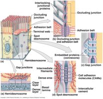
Classification of Epithelia
Epithelia are classified by the number of cell layers and the shape of the cells at the apical surface:
Simple epithelium: Single layer of cells; all cells touch the basement membrane.
Stratified epithelium: Multiple layers; only the deepest layer touches the basement membrane.
Type | Squamous | Cuboidal | Columnar |
|---|---|---|---|
Simple | Flat, thin cells | Cube-shaped cells | Tall, column-like cells |
Stratified | Multiple layers of flat cells | Multiple layers of cube-shaped cells | Multiple layers of column-like cells |
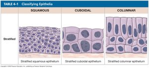
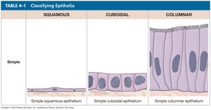
Types of Epithelial Tissue
Simple Squamous Epithelium
Composed of a single layer of flat cells, this epithelium allows for rapid diffusion and filtration. It lines blood vessels (endothelium), body cavities (mesothelium), and alveoli of the lungs.
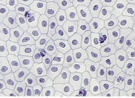
Simple Cuboidal Epithelium
Consists of a single layer of cube-shaped cells with centrally located nuclei. Functions include secretion and absorption. Found in kidney tubules and glandular ducts.
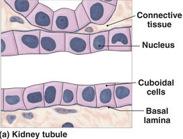
Simple Columnar Epithelium
Made up of a single layer of tall, column-shaped cells, often with microvilli or cilia. Functions in absorption, secretion, and protection. Located in the digestive tract and uterine tubes.
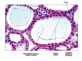
Stratified Squamous Epithelium
Multiple layers of flat cells provide protection against abrasion. Found in the skin, mouth, throat, esophagus, and vagina.
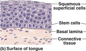
Stratified Cuboidal Epithelium
Two or more layers of cube-shaped cells, providing protection. Found in ducts of sweat glands and mammary glands.
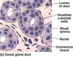
Stratified Columnar Epithelium
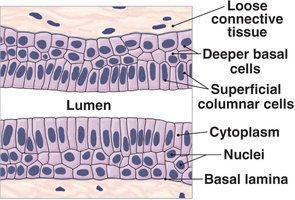
Several layers with columnar cells at the surface. Provides protection and is found in parts of the urethra, anus, and pharynx.
Transitional Epithelium
Specialized for stretching and recoiling, transitional epithelium lines the urinary bladder and ureters. The apical cells appear dome-shaped when relaxed and flattened when stretched.
Pseudostratified Ciliated Columnar Epithelium
Appears layered due to varying positions of nuclei, but all cells touch the basement membrane. Contains cilia and goblet cells. Functions in secretion and movement of mucus. Found in the respiratory tract.
Glandular Epithelium
Glands are classified by where they secrete their products:
Exocrine glands: Secrete into ducts (e.g., sweat, salivary glands).
Endocrine glands: Secrete hormones directly into the bloodstream (e.g., pancreas, adrenal glands).
Modes of secretion include merocrine (exocytosis), apocrine (loss of cytoplasm), and holocrine (cell rupture).
Connective Tissue
Characteristics and Functions
Connective tissue is the most diverse and abundant tissue type. It consists of cells embedded in an extracellular matrix of protein fibers and ground substance. Functions include binding, supporting, protecting, storing energy, and transporting substances.
Major fiber types: Collagen (strength), elastic (stretch), reticular (supportive networks).
Major cell types: Fibroblasts, adipocytes, macrophages, mast cells, and others.
Types of Connective Tissue
Areolar Connective Tissue
Loose arrangement of fibers and cells, providing support and flexibility. Found under the skin and around organs.
Adipose Tissue
Stores fat in large vacuoles, cushions organs, and insulates the body. Located under the skin, around eyes, and in breasts.
Reticular Tissue
Forms a supportive framework for organs such as the liver, spleen, and lymph nodes.
Dense Regular Connective Tissue
Composed of tightly packed collagen fibers aligned in parallel, providing strong attachment. Found in tendons and ligaments.
Elastic Tissue
Dominated by elastic fibers, allowing tissues to stretch and recoil. Found in the walls of large arteries and vertebrae.
Dense Irregular Connective Tissue
Collagen fibers arranged in an irregular pattern, providing strength in multiple directions. Found in the dermis and organ capsules.
Blood
A fluid connective tissue with cells suspended in plasma. Functions in transport, defense, and clotting. Main cell types: erythrocytes (RBCs), leukocytes (WBCs), and platelets.
Lymph
Extracellular fluid that circulates in lymphatic vessels, filtered by lymph nodes, and returned to the bloodstream. Important for immune defense.
Cartilage
Cartilage is a supportive connective tissue with a firm, gel-like matrix. Chondrocytes reside in lacunae. Types include:
Hyaline cartilage: Most common; reduces friction between bones.
Elastic cartilage: Flexible; found in the ear and epiglottis.
Fibrocartilage: Durable; found in intervertebral discs and pubic symphysis.
Bone
Bone is the most rigid connective tissue, with a matrix hardened by calcium salts. Osteocytes are found in lacunae, and the tissue is organized around central canals. Functions include support, protection, and blood cell production.
Muscle and Nervous Tissues (Overview)
Muscle Tissue
Muscle tissue is specialized for contraction and movement. Types include:
Skeletal muscle: Voluntary, striated, multinucleated; attached to bones.
Cardiac muscle: Involuntary, striated, branched; found in the heart.
Smooth muscle: Involuntary, non-striated; found in walls of hollow organs.
Nervous Tissue
Nervous tissue conducts electrical impulses and processes information. Main cell types are neurons (transmit signals) and neuroglia (support and protect neurons). Found in the brain, spinal cord, and nerves.
Membranes
Types of Membranes
Membranes are physical barriers composed of epithelial and connective tissues. Types include:
Mucous membranes: Line passageways open to the exterior (e.g., digestive, respiratory tracts).
Serous membranes: Line internal cavities; secrete serous fluid to reduce friction.
Cutaneous membrane: The skin; thick, waterproof, and dry.
Synovial membranes: Line joint cavities; produce synovial fluid for lubrication.
