 Back
BackThe Tissue Level of Organization: Structure and Function of Body Tissues
Study Guide - Smart Notes
 Tailored notes based on your materials, expanded with key definitions, examples, and context.
The Tissue Level of Organization
Introduction to Tissues and Histology
Tissues are collections of specialized cells and cell products that perform specific, limited functions. The study of tissues is known as histology. Understanding tissues is essential for comprehending how organs and organ systems function in the human body.
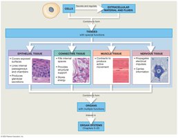
Epithelial Tissue
Overview and Characteristics
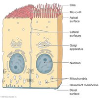
Epithelial tissue includes layers of cells that cover internal or external surfaces and glands that produce secretions. Key characteristics include:
Cells bound closely together
Free (apical) surface exposed to the environment
Attachment to underlying connective tissue by a basement membrane
Avascular (lacking blood vessels)
Continual replacement or regeneration of cells
Locations and Functions
Epithelia cover both external and internal body surfaces, forming selective barriers and lining internal cavities and passageways. Major functions include:
Physical protection
Control of permeability
Provision of sensation
Production of specialized secretions (glandular epithelium)
Glandular Epithelium
Gland cells are classified by where their secretions are discharged:
Exocrine glands: Secrete onto epithelial surfaces via ducts
Endocrine glands: Release hormones into interstitial fluid and blood (ductless)
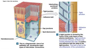
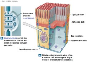
Intercellular Connections (Cell Junctions)
Cell junctions allow firm attachment between epithelial cells and to the basement membrane. Major types include:
Tight junctions: Prevent passage of substances between cells
Adhesion belts: Reinforce tight junctions
Gap junctions: Permit communication between cells
Desmosomes: Provide strong attachment between cells
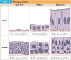
Classification of Epithelia
Epithelia are classified by the number of cell layers and the shape of the cells at the apical surface:
Squamous | Cuboidal | Columnar | |
|---|---|---|---|
Simple | Simple squamous epithelium | Simple cuboidal epithelium | Simple columnar epithelium |
Stratified | Stratified squamous epithelium | Stratified cuboidal epithelium | Stratified columnar epithelium |
Examples of Epithelial Types
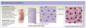
Simple squamous epithelium: Thin, flat cells; found in alveoli of lungs, lining of heart and blood vessels. Function: diffusion and filtration.
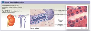
Simple cuboidal epithelium: Cube-shaped cells; found in kidney tubules, glands. Function: secretion and absorption.
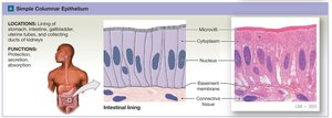
Simple columnar epithelium: Tall, column-like cells; found in digestive tract lining. Function: absorption and secretion.
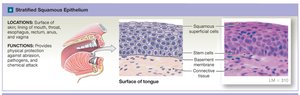
Stratified squamous epithelium: Multiple layers; found in skin, mouth, esophagus. Function: protection.
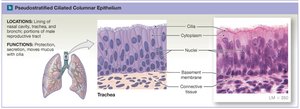
Pseudostratified columnar epithelium: Appears layered but all cells touch basement membrane; found in respiratory tract. Function: protection, secretion.
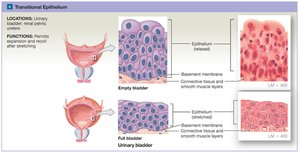
Transitional epithelium: Appearance changes with stretching; found in urinary bladder. Function: permits expansion and recoil.
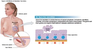
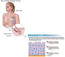
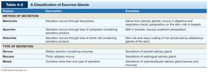
Modes of Exocrine Secretion
Exocrine glands secrete by three main methods:
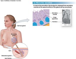
Merocrine secretion: Product released by exocytosis (e.g., salivary glands)
Apocrine secretion: Involves loss of cytoplasm with secretion (e.g., mammary glands)
Holocrine secretion: Entire cell bursts, releasing contents (e.g., sebaceous glands)
Method of Secretion | Description | Examples |
|---|---|---|
Merocrine | Secretion occurs through exocytosis | Saliva from salivary glands, sweat |
Apocrine | Secretion occurs through loss of cytoplasm | Milk in breasts, underarm perspiration |
Holocrine | Secretion occurs through loss of entire cell | Sebaceous (oil) glands |
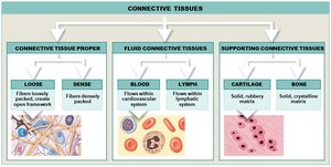
Connective Tissue
Overview and Characteristics
Connective tissue is the most diverse tissue type, providing a structural framework, support, and protection for other tissues. It consists of specialized cells, extracellular protein fibers, and ground substance (fluid).
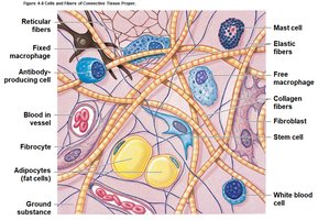
Cells and Fibers of Connective Tissue Proper
Fibroblasts: Produce fibers and ground substance
Fibrocytes: Maintain fibers
Macrophages: Phagocytize pathogens and debris
Adipocytes: Store fat
Mast cells: Release histamine and heparin during inflammation
Types of fibers:
Collagen fibers: Strong, flexible, most common
Elastic fibers: Stretch and return to original length
Reticular fibers: Form supportive networks
Types of Connective Tissue Proper
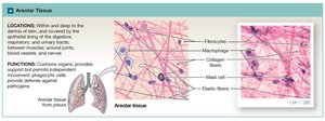
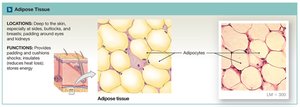
Loose connective tissue: More ground substance, fewer fibers; supports epithelia, fills spaces, stores lipids
Areolar tissue: Cushions organs, provides support
Adipose tissue: Stores energy, insulates, cushions
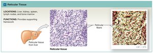
Reticular tissue: Provides supporting framework
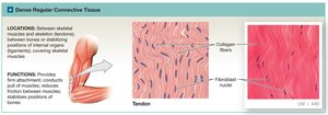
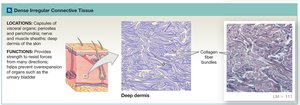
Dense connective tissue: More fibers, less ground substance; resists tension
Dense regular connective tissue: Parallel collagen fibers; tendons, ligaments
Dense irregular connective tissue: Interwoven fibers; dermis, organ capsules
Fluid and Supporting Connective Tissues
Fluid connective tissues: Blood and lymph; transport cells and dissolved materials
Supporting connective tissues: Cartilage and bone; provide strong framework
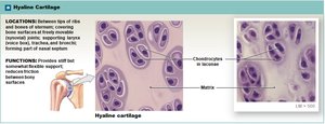
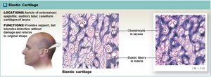
Cartilage
Hyaline cartilage: Most common; closely packed collagen fibers; found in joints
Elastic cartilage: Contains elastic fibers; flexible; found in ear
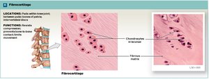
Fibrocartilage: Densely woven collagen fibers; durable; found in intervertebral discs
Bone (Osseous Tissue)
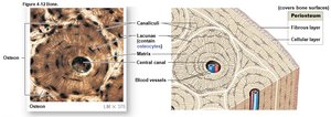
Bone tissue has a hard matrix of calcium compounds and flexible collagen fibers. Osteocytes are arranged around central canals and communicate via canaliculi. The periosteum covers bone surfaces except at joints.

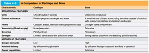
Feature | Cartilage | Bone |
|---|---|---|
Cells | Chondrocytes in lacunae | Osteocytes in lacunae |
Ground substance | Chondroitin sulfate gel | Small volume of liquid with calcium salts |
Fibers | Collagen, elastic, reticular | Collagen fibers (predominate) |
Vascularity | Avascular | Vascular |
Repair | Limited | Extensive |
Tissue Membranes
Types of Tissue Membranes
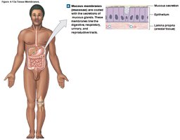
Tissue membranes are physical barriers that line or cover body surfaces. They consist of an epithelium and supporting connective tissue. Types include:
Mucous membranes: Line passageways open to exterior; kept moist by secretions
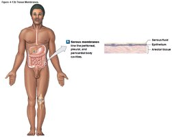
Serous membranes: Line sealed internal cavities; secrete serous fluid to reduce friction
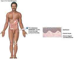
Cutaneous membrane: Skin; thick, waterproof, dry
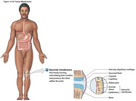
Synovial membranes: Line joint cavities; produce synovial fluid for lubrication
Muscle Tissue
Types and Functions
Muscle tissue is specialized for contraction and movement. There are three types:
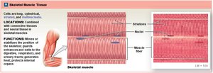
Skeletal muscle: Voluntary, striated, moves skeleton
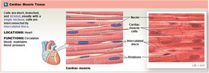
Cardiac muscle: Involuntary, striated, found in heart, intercalated discs for coordination
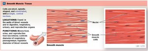
Smooth muscle: Involuntary, nonstriated, found in walls of organs
Nervous Tissue
Structure and Function
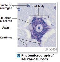
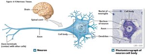
Nervous tissue is specialized for transmitting electrical impulses. It is concentrated in the brain and spinal cord and is essential for sensing the environment and controlling responses.
Neurons: Nerve cells that transmit signals
Neuroglia: Supporting cells that protect and nourish neurons
Tissue Injury and Repair
Inflammation and Regeneration
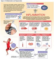
Tissues respond to injury through inflammation (first response) and regeneration (repair). Inflammation involves swelling, heat, redness, and pain, triggered by trauma or infection. Mast cells release chemicals that increase blood flow and attract immune cells. Regeneration replaces damaged tissue; some tissues regenerate well (epithelia, connective), others poorly (cardiac, neural).
Aging and Tissue Changes
With age, tissue repair slows, and the risk of cancer increases. Epithelia thin, connective tissues become fragile, bones brittle, and cartilage less resilient. Cancer rates rise due to accumulated mutations from environmental exposures.
