 Back
BackThe Tissue Level of Organization: Structure and Function of Human Tissues
Study Guide - Smart Notes
 Tailored notes based on your materials, expanded with key definitions, examples, and context.
The Tissue Level of Organization
Introduction to Tissues
Tissues are collections of specialized cells and cell products that perform specific functions. The study of tissues is known as histology. Tissues combine to form organs, such as the heart and lungs, each with specialized roles in the body.
Types of Tissues
Epithelial Tissue: Covers exposed surfaces, lines internal passageways, and forms glands.
Connective Tissue: Fills internal spaces, supports other tissues, transports materials, and stores energy.
Muscle Tissue: Specialized for contraction, found in skeletal muscles, the heart, and walls of hollow organs.
Nervous Tissue: Carries electrical signals throughout the body.
Epithelial Tissue
Characteristics of Epithelia
Epithelial tissues exhibit several key characteristics:
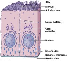
Polarity: Distinct apical (top) and basal (bottom) surfaces.
Cellularity: Composed almost entirely of tightly packed cells connected by cell junctions.
Attachment: Bound to a basement membrane.
Avascularity: Lacks blood vessels; nutrients diffuse from underlying tissues.
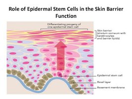
Regeneration: High capacity for renewal via stem cells.
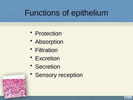
Functions of Epithelium
Protection
Absorption
Filtration
Excretion
Secretion
Sensory reception
Maintaining Epithelial Integrity
Intercellular Connections: Cell adhesion molecules (CAMs), proteoglycans, and specialized junctions (gap, tight, desmosomes).
Attachment to Basement Membrane: Provides support and anchorage.
Maintenance and Repair: Stem cells near the basement membrane ensure renewal.
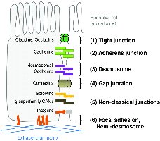
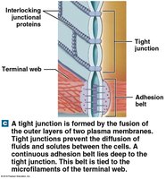
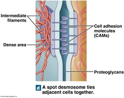
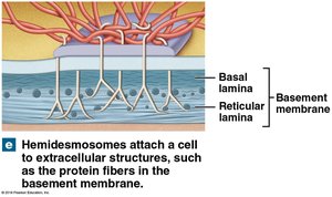
Types of Cell Junctions
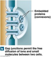
Gap Junctions: Allow rapid communication and passage of ions/small molecules.
Tight Junctions: Prevent passage of water and solutes between cells.
Desmosomes: Provide mechanical strength by tying cells together.
Hemidesmosomes: Anchor cells to the basement membrane.
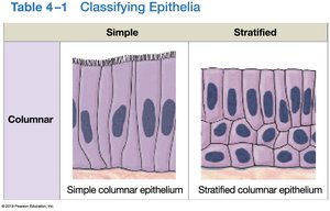
Classification of Epithelia
Epithelia are classified by cell shape and number of layers:
Shapes: Squamous (flat), Cuboidal (cube-shaped), Columnar (tall/rectangular)
Layers: Simple (one layer), Stratified (multiple layers)
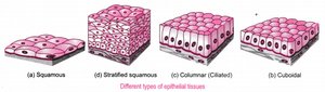
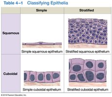
Examples of Epithelial Types
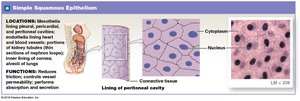
Simple Squamous: Absorption/diffusion (e.g., alveoli, blood vessels)
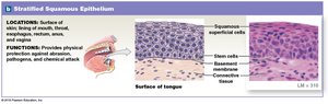
Stratified Squamous: Protection against abrasion (e.g., skin, mouth)
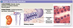
Simple Cuboidal: Secretion/absorption (e.g., kidney tubules)
Stratified Cuboidal: Protection (rare; sweat glands)
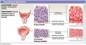
Transitional: Stretches (urinary bladder)
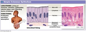
Simple Columnar: Absorption/secretion (digestive tract)
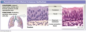
Pseudostratified Columnar: Protection, movement of mucus (respiratory tract)
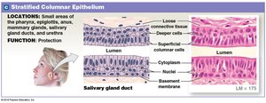
Stratified Columnar: Protection (rare; pharynx, anus)
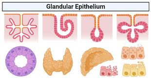
Glandular Epithelia
Glands are collections of epithelial cells that produce secretions. They are classified as:
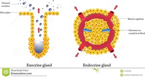
Endocrine Glands: Release hormones into the bloodstream (ductless).
Exocrine Glands: Release secretions onto epithelial surfaces via ducts.
Methods of Secretion
Merocrine: Released by exocytosis (e.g., sweat glands).
Apocrine: Released by shedding cytoplasm (e.g., mammary glands).
Holocrine: Released by cell bursting (e.g., sebaceous glands).
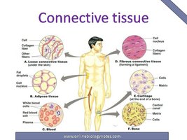
Connective Tissue
General Features
Connective tissues are the most diverse and abundant tissues, providing support, protection, and integration of all body parts. They consist of specialized cells, extracellular protein fibers, and ground substance, which together form the matrix.
Functions of Connective Tissue
Structural framework for the body
Transport of fluids and dissolved materials
Protection of organs
Support, surround, and interconnect other tissues
Energy storage (triglycerides)
Defense against microorganisms
Categories of Connective Tissue
Connective Tissue Proper: Connects and protects (e.g., adipose, tendons)
Fluid Connective Tissues: Transport (e.g., blood, lymph)
Supporting Connective Tissues: Structural strength (e.g., cartilage, bone)
Cells of Connective Tissue Proper
Fibroblasts: Produce fibers and ground substance
Fibrocytes: Maintain fibers
Adipocytes: Store fat
Mesenchymal Cells: Stem cells for repair
Macrophages: Phagocytize pathogens and debris
Mast Cells: Release histamine/heparin (inflammation)
Lymphocytes: Immune response
Microphages: Phagocytic blood cells
Melanocytes: Synthesize melanin
Connective Tissue Fibers
Collagen Fibers: Strong, resist tension (tendons, ligaments)
Reticular Fibers: Form supportive networks (stroma)
Elastic Fibers: Stretch and return to original length (elastic ligaments)
Types of Connective Tissue Proper
Loose Connective Tissue: More ground substance, fewer fibers (e.g., areolar, adipose, reticular)
Dense Connective Tissue: More fibers, less ground substance (e.g., dense regular, dense irregular, elastic)
Fluid Connective Tissues
Blood: Contains plasma, red blood cells, white blood cells, and platelets
Lymph: Forms from interstitial fluid, monitored by immune system
Supporting Connective Tissues
Cartilage: Shock absorption, flexible support (hyaline, elastic, fibrocartilage)
Bone (Osseous Tissue): Weight support, rigid due to calcium salts and collagen fibers
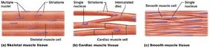
Muscle Tissue
Types of Muscle Tissue
Skeletal Muscle: Voluntary, striated, moves the skeleton
Cardiac Muscle: Involuntary, striated, found only in the heart
Smooth Muscle: Involuntary, non-striated, found in walls of hollow organs
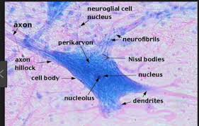
Nervous Tissue
Structure and Function
Nervous tissue is specialized for conducting electrical impulses and is concentrated in the brain and spinal cord. It consists of two main cell types:
Neurons: Transmit electrical signals
Neuroglia: Support and protect neurons
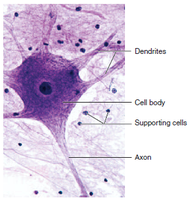
Parts of a Neuron
Cell Body: Contains nucleus and organelles
Dendrites: Receive incoming signals
Axon: Transmits signals to other cells
Tissue Membranes
Types of Membranes
Mucous Membranes: Line passageways with external openings (digestive, respiratory tracts)
Serous Membranes: Line internal cavities, secrete serous fluid
Cutaneous Membrane: Skin, covers body surface
Synovial Membranes: Line joint cavities, produce synovial fluid
Tissue Response to Injury and Aging
Inflammation and Regeneration
Inflammation: Triggered by trauma or infection, involves release of chemicals and activation of immune cells
Regeneration: Replacement of damaged tissue; varies by tissue type (epithelia and connective regenerate well, muscle and nervous poorly)
Aging and Tissue Structure
Decreased speed and effectiveness of tissue repair
Thinner epithelia, fragile connective tissues, increased risk of disease
Increased cancer incidence with age
Additional info: This guide covers the essential concepts of tissue structure and function, as outlined in Chapter 4 of a standard Anatomy & Physiology curriculum. It is suitable for exam preparation and foundational understanding for further study in human biology and health sciences.
