 Back
BackThe Urinary System: Structure and Function
Study Guide - Smart Notes
 Tailored notes based on your materials, expanded with key definitions, examples, and context.
The Urinary System
Introduction to the Urinary System
The urinary system is essential for maintaining homeostasis by removing metabolic wastes, regulating blood volume and pressure, controlling plasma ion concentrations, and contributing to pH balance. It consists of paired kidneys, ureters, the urinary bladder, and the urethra.
Removes metabolic wastes via urine
Osmoregulation: Regulates blood volume, blood pressure, and plasma ion concentrations
pH homeostasis: Controls loss of hydrogen and bicarbonate ions
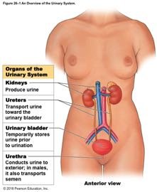
Organs of the Urinary System
Major Components and Their Functions
Kidneys: Produce urine by filtering blood and removing wastes
Ureters: Transport urine from kidneys to urinary bladder
Urinary bladder: Temporarily stores urine prior to elimination
Urethra: Conducts urine to the exterior; in males, also transports semen
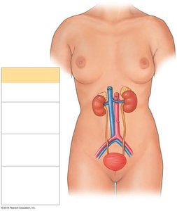
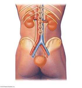
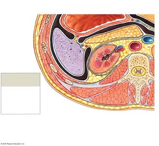
Anatomical Position and Protection of the Kidneys
The kidneys are retroperitoneal organs located on either side of the vertebral column. The left kidney is slightly superior to the right. Each kidney is protected and stabilized by three layers:
Fibrous capsule: Collagen fibers covering the outer surface
Perinephric fat: Thick layer of adipose tissue surrounding the capsule
Renal fascia: Dense, fibrous outer layer anchoring the kidney
Gross and Internal Anatomy of the Kidney
External and Internal Structures
The kidney has a distinct outer cortex and inner medulla. The medulla contains renal pyramids, which are separated by renal columns. The renal pelvis collects urine from the major calyces and channels it into the ureter.
Renal cortex: Outer region containing nephrons
Renal medulla: Inner region with renal pyramids
Renal pelvis: Funnel-shaped structure collecting urine
Hilum: Entry/exit site for renal artery, vein, and ureter
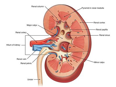

Pathway of Urine Flow
Urine is produced in the nephrons, passes through the collecting ducts, and drains into minor calyces, then major calyces, the renal pelvis, and finally the ureter.
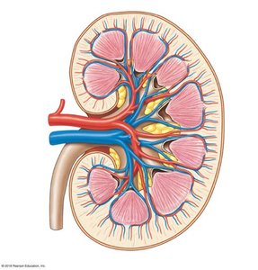
Blood Supply and Innervation of the Kidneys
Renal Circulation
The kidneys receive about 20–25% of cardiac output. Blood enters via the renal artery, branches into smaller arteries, and is filtered in the glomerulus. Venous blood returns via the renal vein.
Renal artery → segmental arteries → interlobar arteries → arcuate arteries → cortical radiate arteries → afferent arterioles → glomerulus
Efferent arterioles → peritubular capillaries/vasa recta → cortical radiate veins → arcuate veins → interlobar veins → renal vein
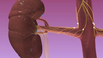
Renal Nerves
Sympathetic innervation regulates blood flow and urine formation by adjusting the diameter of renal arterioles and stimulating renin release.
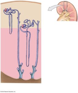
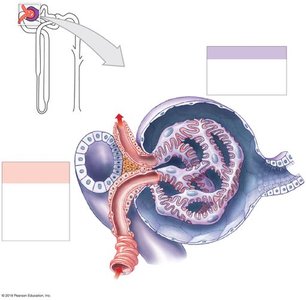
Microscopic Structure: The Nephron
Nephron Anatomy and Types
Nephrons are the functional units of the kidney, each consisting of a renal corpuscle and a renal tubule. There are two main types:
Cortical nephrons: 85% of nephrons, located mostly in the cortex, with short loops of Henle
Juxtamedullary nephrons: 15% of nephrons, with long loops extending deep into the medulla, crucial for concentrating urine

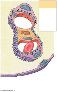
Renal Corpuscle
The renal corpuscle consists of the glomerulus (a capillary network) and the glomerular (Bowman's) capsule. Filtration occurs here, producing a protein-free filtrate.
Podocytes: Specialized cells with foot processes forming filtration slits
Filtration membrane: Composed of fenestrated endothelium, basement membrane, and podocyte foot processes
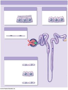
Renal Tubule and Collecting System
The renal tubule consists of the proximal convoluted tubule (PCT), nephron loop (loop of Henle), and distal convoluted tubule (DCT). The collecting system receives fluid from multiple nephrons and transports it to the renal pelvis.
PCT: Reabsorbs water, ions, and nutrients
Nephron loop: Establishes osmotic gradient for water reabsorption
DCT: Secretion and selective reabsorption under hormonal control
Collecting duct: Final adjustments to urine composition and volume

Summary Table: Main Structures of the Kidney
Structure | Function |
|---|---|
Renal Cortex | Contains renal corpuscles and most of the nephron tubules |
Renal Medulla | Contains renal pyramids and nephron loops |
Renal Pelvis | Collects urine from calyces and channels it to the ureter |
Hilum | Entry/exit for renal artery, vein, and ureter |
Ureter | Transports urine to the bladder |
Key Terms and Concepts
Osmoregulation: Regulation of water and solute concentrations in the blood
Filtration: Movement of water and solutes from blood into the nephron
Reabsorption: Movement of substances from filtrate back into the blood
Secretion: Movement of substances from blood into the filtrate
Renin: Enzyme involved in blood pressure regulation
ADH (Antidiuretic Hormone): Regulates water reabsorption in the collecting ducts
