 Back
BackThe Urinary System: Structure, Function, and Clinical Relevance
Study Guide - Smart Notes
 Tailored notes based on your materials, expanded with key definitions, examples, and context.
The Urinary System
Overview and Major Components
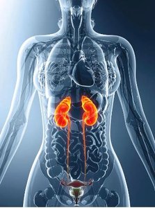
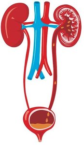
The urinary system is essential for the removal of waste products and the regulation of blood composition. It consists of the kidneys, ureters, urinary bladder, and urethra.
Kidneys: Organs that produce urine by filtering blood.
Urinary tract: Includes paired ureters, the urinary bladder, and the urethra, which eliminate urine from the body.
Urination (micturition): The process of expelling urine via contraction of the bladder.
Functions of the Urinary System
The urinary system performs three primary functions:
Excretion: Removal of organic wastes from body fluids.
Homeostatic regulation: Maintains blood plasma volume and solute concentration.
Elimination: Discharge of waste products from the body.
Homeostatic Functions
Regulation of blood volume and blood pressure: Adjusts water loss and releases erythropoietin and renin.
Regulation of plasma ion concentrations: Controls sodium, potassium, chloride, and calcium ions.
Stabilization of blood pH: Manages hydrogen and bicarbonate ion loss.
Conservation of nutrients: Prevents excretion of valuable nutrients while eliminating wastes.
Assistance in detoxification: Supports the liver in detoxifying poisons.
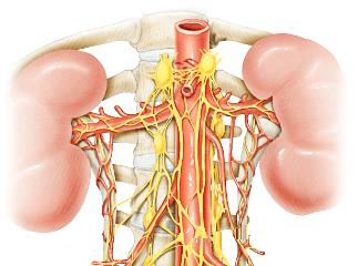
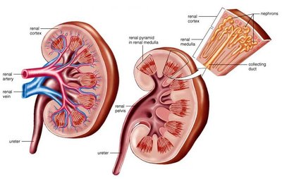
The Kidneys
Position, Protection, and Structure
The kidneys are retroperitoneal organs stabilized by renal fascia, contact with adjacent organs, a fibrous capsule, and a perinephric fat capsule.
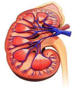
Renal Cortex: The superficial, granular region in contact with the renal capsule.
Renal Pyramids: Triangular structures in the medulla, with bases aligned along the cortex and tips (renal papillae) projecting into the renal sinus.
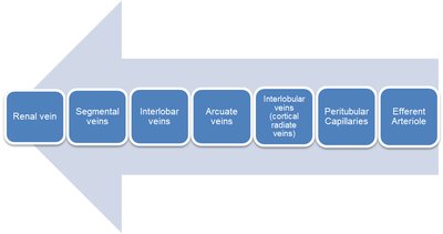
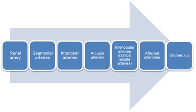
Blood Supply to the Kidneys
The kidneys receive 20–25% of cardiac output, with about 1200 mL of blood flowing through them each minute. Blood enters via the renal arteries and leaves via the renal veins. 
Renal Innervation
Renal plexus: Nerves enter at the hilus and innervate kidneys and ureters.
Sympathetic innervation: Adjusts urine formation rate by altering blood flow and pressure, and stimulates renin release.
Anatomy of the Kidney: Sectional View
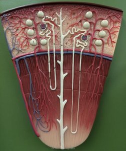
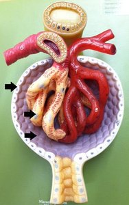
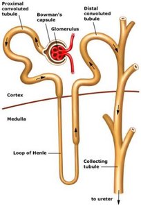
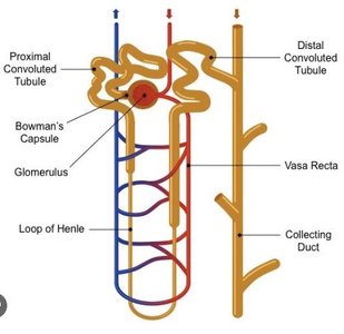
Nephrons
Nephrons are the functional units of the kidney, responsible for urine production.
Cortical nephrons: ~85% of nephrons, located mostly in the cortex, with short loops of Henle.
Juxtamedullary nephrons: ~15% of nephrons, loops extend deep into the medulla, connect to vasa recta.
Nephron Structure
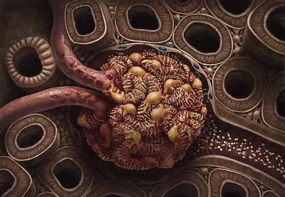
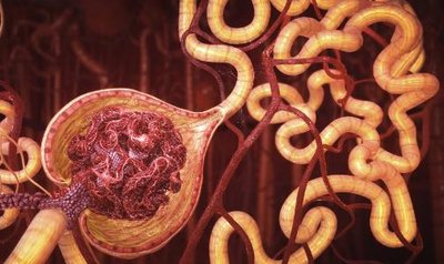
Renal corpuscle: Spherical structure with the glomerular (Bowman's) capsule and glomerulus.
Renal tubule: Long passageway beginning at the corpuscle.

Renal Corpuscle
Glomerular capsule: Lined by simple squamous epithelium, continuous with visceral epithelium covering glomerular capillaries, separated by capsular space.
Glomerulus: Extensive capillary bed; blood enters via afferent arteriole and leaves via efferent arteriole.
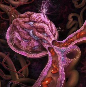
Filtration in the Glomerulus
Filtration is a passive process driven by blood pressure, forcing water and solutes out of glomerular capillaries into the capsular space.
Produces protein-free filtrate similar to blood plasma.
Larger solutes (e.g., plasma proteins) remain in the blood.
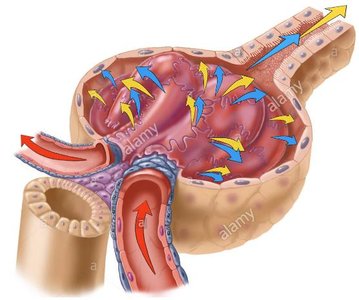
Solutes Entering Capsular Space
Metabolic wastes
Excess ions
Glucose, free fatty acids, amino acids, vitamins
Reabsorption
Useful materials are reabsorbed from the filtrate, primarily in the proximal convoluted tubule (PCT), before leaving the kidneys. 
Segments of the Renal Tubule
Proximal convoluted tubule (PCT): Located in cortex, first segment after corpuscle.
Nephron loop (Loop of Henle): U-shaped tube extending into medulla.
Distal convoluted tubule (DCT): Located in cortex, third segment, smaller diameter than PCT.
Functions of the Renal Tubule
Reabsorb useful organic nutrients from filtrate.
Reabsorb more than 90% of water in filtrate.
Secrete waste products not filtered at the glomerulus.
Proximal Convoluted Tubule (PCT)
First segment of renal tubule.
Entrance lies opposite the connection of afferent and efferent arterioles.

Nephron Loop (Loop of Henle)
Descending limb: Fluid flows toward renal pelvis.
Ascending limb: Fluid flows toward renal cortex.
Each limb contains thick and thin segments.

Distal Convoluted Tubule (DCT)
Third segment of renal tubule.
Initial portion passes between afferent and efferent arterioles.
Smaller diameter than PCT.
Processes at the DCT
Active secretion of ions, acids, drugs, and toxins.
Selective reabsorption of Na+ and Ca2+ ions.
Selective reabsorption of water, concentrating tubular fluid.
Collecting Ducts
Collecting ducts adjust fluid composition and determine the final osmotic concentration and volume of urine.
Receive tubular fluid from DCT of many nephrons.
Begin in cortex, descend into medulla, and drain into minor calyx.


Juxtaglomerular Complex
The juxtaglomerular complex is an endocrine structure that secretes erythropoietin and renin. It is formed by the macula densa and juxtaglomerular cells.
Macula Densa
Epithelial cells of DCT near the renal corpuscle.
Tall cells with densely clustered nuclei.
Juxtaglomerular Cells
Smooth muscle fibers in the wall of the afferent arteriole.
Form the juxtaglomerular complex with macula densa.
Kidney Diseases and Disorders
Chronic Kidney Disease (CKD)
High blood pressure damages glomeruli, leading to kidney function decline.
Diabetes is a major cause.
Treatment: dialysis, organ transplant.
Nephrolithiasis (Kidney Stones)
Minerals crystallize in kidneys, forming stones.
Symptoms: pain, rarely cause long-term problems.
Treatment: increased water intake, passing stones, medical procedures for large stones.
Glomerulonephritis
Inflammation of glomeruli due to infection, drugs, or congenital abnormalities.
Symptoms: hematuria (pinkish urine).
Treatment: dietary changes, stop smoking, lower BP, often resolves itself.
Polycystic Kidney Disease (PKD)
Genetic disorder causing growth of fluid-filled sacs (cysts).
Symptoms: high BP, back/side pain, kidney failure by age 60 in many cases.
Treatment: BP control, pain relief, cyst removal, kidney transplant.
Urinary Tract Infection (UTI)
Bacterial infection of any part of the urinary system.
More serious when kidneys are involved.
More common in women; symptoms include pelvic pain, urge to urinate, pain with urination, back/side pain, nausea, vomiting, fever.
Treatment: antibiotics.
Pyelonephritis (Kidney Infection)
Inflammation of the kidney due to bacterial infection.
Usually begins in the urethra and travels upward.
Treatment: antibiotics, hospitalization.
Summary Table: Major Kidney Functions
Function | Description |
|---|---|
Excretion | Removal of organic wastes from body fluids |
Homeostatic Regulation | Regulation of blood volume, plasma ion concentration, and pH |
Elimination | Discharge of waste products |
Detoxification | Assists liver in detoxifying poisons |
Summary Table: Nephron Segments and Functions
Segment | Main Function |
|---|---|
Renal Corpuscle | Filtration of blood |
Proximal Convoluted Tubule (PCT) | Reabsorption of nutrients, ions, and water |
Loop of Henle | Concentration of urine |
Distal Convoluted Tubule (DCT) | Secretion and selective reabsorption |
Collecting Duct | Final adjustment of urine composition |
