 Back
BackThe Urinary System: Structure, Function, and Physiology
Study Guide - Smart Notes
 Tailored notes based on your materials, expanded with key definitions, examples, and context.
The Urinary System: Structure and Function
Overview of the Urinary System
The urinary system is essential for maintaining the body's internal environment by regulating water, electrolyte, and acid-base balance, as well as removing metabolic wastes. It consists of the kidneys, ureters, urinary bladder, and urethra.
Kidneys: Major excretory organs responsible for filtering blood and forming urine.
Ureters: Transport urine from the kidneys to the urinary bladder.
Urinary bladder: Temporarily stores urine until excretion.
Urethra: Conducts urine from the bladder to the outside of the body.
Kidney Structure and Function
Internal Anatomy of the Kidney
The kidney is divided into the renal cortex and renal medulla. The medulla contains cone-shaped renal pyramids separated by renal columns. The tip of each pyramid, called the papilla, releases urine into minor calyces, which then drain into major calyces and finally the renal pelvis, a funnel-shaped tube continuous with the ureter.
Blood and Nerve Supply
The kidneys receive about 25% of cardiac output via the renal arteries, reflecting their critical role in filtering blood. The nerve supply is primarily sympathetic, regulating blood flow and filtration rate.
Nephrons: The Functional Units
Structure of the Nephron
Each kidney contains over one million nephrons, which are responsible for urine formation. Each nephron consists of a renal corpuscle and a renal tubule.
Renal corpuscle: Includes the glomerulus (a tuft of capillaries) and the glomerular (Bowman's) capsule.
Renal tubule: Subdivided into the proximal convoluted tubule (PCT), nephron loop (loop of Henle), and distal convoluted tubule (DCT).
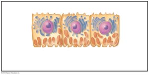
Renal Tubule Cell Types
The renal tubule is lined with specialized epithelial cells that facilitate reabsorption and secretion. For example, the PCT contains cuboidal cells with dense microvilli for efficient reabsorption.
Collecting Ducts
Collecting ducts receive filtrate from multiple nephrons and play a key role in water and electrolyte balance. They contain principal cells (regulate water and Na+ balance) and intercalated cells (regulate acid-base balance).
Classes of Nephrons
Cortical nephrons: Make up 85% of nephrons; located mostly in the cortex.
Juxtamedullary nephrons: Have long loops that extend deep into the medulla; crucial for concentrating urine.
Nephron Capillary Beds and Juxtaglomerular Complex
Capillary Beds
Glomerulus: Specialized for filtration; fed and drained by arterioles.
Peritubular capillaries: Adapted for absorption; surround the renal tubule.
Vasa recta: Long, thin-walled vessels associated with juxtamedullary nephrons; maintain medullary osmotic gradient.
Juxtaglomerular Complex (JGC)
The JGC regulates filtrate formation and blood pressure. It includes macula densa cells (chemoreceptors for NaCl), granular cells (mechanoreceptors that secrete renin), and extraglomerular mesangial cells (signal transmission).
Mechanisms of Urine Formation
Three Main Processes
Glomerular filtration: Passive process driven by hydrostatic pressure; produces cell- and protein-free filtrate.
Tubular reabsorption: Selective return of substances from filtrate to blood; includes both active and passive mechanisms.
Tubular secretion: Selective movement of substances from blood into filtrate for excretion.
Filtration Membrane
The filtration membrane consists of three layers: fenestrated endothelium, basement membrane, and podocyte foot processes. It allows passage of water and small solutes but restricts proteins and cells.
Pressures Affecting Filtration
Outward pressure: Hydrostatic pressure in glomerular capillaries (~55 mm Hg) promotes filtration.
Inward pressures: Hydrostatic pressure in the capsular space (15 mm Hg) and colloid osmotic pressure in capillaries (30 mm Hg) oppose filtration.
Net Filtration Pressure (NFP):
Glomerular Filtration Rate (GFR)
GFR is the volume of filtrate formed per minute by both kidneys (normal: 120–125 ml/min). It is directly proportional to NFP, filtration surface area, and membrane permeability.
Regulation of Glomerular Filtration
Intrinsic Controls (Renal Autoregulation)
Myogenic mechanism: Smooth muscle response to pressure changes in afferent arterioles.
Tubuloglomerular feedback: Macula densa cells sense NaCl and adjust arteriole diameter to regulate GFR.
Extrinsic Controls
Sympathetic nervous system: Reduces GFR during low blood pressure by constricting afferent arterioles.
Renin-angiotensin-aldosterone system: Increases blood pressure and GFR via hormonal pathways.
Tubular Reabsorption and Secretion
Reabsorption Routes
Transcellular route: Through tubule cells.
Paracellular route: Between tubule cells (limited by tight junctions).
Transport Maximum (Tm)
Each substance has a Tm, reflecting the number of available transport proteins. When exceeded, excess is excreted in urine (e.g., glucose in diabetes).
Reabsorptive Capabilities by Segment
PCT: Most reabsorption occurs here (all nutrients, 65% Na+ and water).
Nephron loop: Descending limb reabsorbs water; ascending limb reabsorbs solutes.
DCT and collecting duct: Reabsorption is hormonally regulated (ADH, aldosterone, atrial natriuretic peptide, parathyroid hormone).
Summary of Tubular Reabsorption and Secretion
Tubular Secretion
Tubular secretion removes substances such as K+, H+, NH4+, creatinine, and drugs from blood into filtrate. It is essential for disposing of bound substances, eliminating wastes, and regulating blood pH.
Regulation of Urine Concentration and Volume
Osmolality and Countercurrent Mechanisms
Osmolality is the number of solute particles per liter of water. The kidneys maintain plasma osmolality at ~300 mOsm using countercurrent mechanisms, which establish and maintain an osmotic gradient in the medulla, allowing for the production of dilute or concentrated urine.
Countercurrent multiplier: Nephron loop creates the gradient.
Countercurrent exchanger: Vasa recta preserves the gradient.
Renal Clearance
Renal clearance is the volume of plasma cleared of a substance per minute. It is calculated as:
C: Renal clearance rate (ml/min)
U: Concentration of substance in urine (mg/ml)
V: Urine flow rate (ml/min)
P: Concentration in plasma (mg/ml)
Inulin is used as a standard because it is freely filtered and neither reabsorbed nor secreted.
Physical and Chemical Characteristics of Urine
Physical Characteristics
Color: Clear, pale to deep yellow (urochrome pigment).
Odor: Slightly aromatic; may develop ammonia odor upon standing.
pH: Slightly acidic (average pH 6; range 4.5–8.0).
Specific gravity: 1.001–1.035, depending on solute concentration.
Chemical Composition
95% water, 5% solutes (mainly nitrogenous wastes: urea, uric acid, creatinine).
Other solutes: Na+, K+, PO43–, SO42–, Ca2+, Mg2+, HCO3–.
Abnormal components may indicate pathology (e.g., proteins, blood cells, bile pigments).
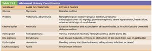
Abnormal Urinary Constituents
Substance | Name of Condition | Possible Causes |
|---|---|---|
Glucose | Glycosuria | Diabetes mellitus |
Proteins | Proteinuria, albuminuria | Nonpathological: excessive exertion, pregnancy; Pathological: kidney damage, hypertension, heart failure |
Ketone bodies | Ketonuria | Starvation, diabetes mellitus |
Hemoglobin | Hemoglobinuria | Hemolytic anemia, transfusion reactions, burns |
Bile pigments | Bilirubinuria | Liver disease, obstruction of bile ducts |
Erythrocytes (RBCs) | Hematuria | Trauma, infection, kidney stones |
Leukocytes (WBCs) | Pyuria | Urinary tract infection |
Urine Transport, Storage, and Elimination
Ureters
Ureters are muscular tubes that convey urine from the kidneys to the bladder. They have three layers: mucosa (transitional epithelium), muscularis (smooth muscle), and adventitia (connective tissue). Peristaltic contractions propel urine toward the bladder.
Urinary Bladder
The bladder is a muscular sac for temporary urine storage. It has a mucosal lining, a thick detrusor muscle, and an outer adventitia. The trigone region is prone to infection. The bladder can expand significantly without a large increase in internal pressure.
Urethra
The urethra is a muscular tube that drains urine from the bladder. It is lined by different types of epithelium along its length and contains internal (involuntary) and external (voluntary) sphincters. The female urethra is short (3–4 cm), while the male urethra is longer and divided into prostatic, membranous, and spongy regions.
Micturition (Urination)
Control of Micturition
Micturition involves the coordinated contraction of the detrusor muscle (by the autonomic nervous system), opening of the internal urethral sphincter (autonomic), and relaxation of the external urethral sphincter (somatic). Reflexive urination in infants is replaced by voluntary control as the nervous system matures.
Pontine storage center: Inhibits urination.
Pontine micturition center: Promotes urination.
