 Back
BackThe Urinary System: Structure, Function, and Regulation
Study Guide - Smart Notes
 Tailored notes based on your materials, expanded with key definitions, examples, and context.
The Urinary System
Introduction to the Urinary System
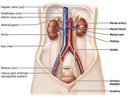
The urinary system is essential for maintaining the body's internal environment by regulating water, solute concentrations, and removing metabolic wastes. It consists of the kidneys, ureters, urinary bladder, and urethra.
Kidneys: Filter blood, regulate fluid and electrolyte balance, and produce urine.
Ureters: Transport urine from kidneys to the bladder.
Urinary bladder: Temporarily stores urine.
Urethra: Conducts urine out of the body.
Kidney Structure and Blood Supply
Location and External Anatomy
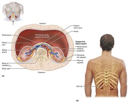
The kidneys are retroperitoneal, bean-shaped organs located in the superior lumbar region. The right kidney is slightly lower due to the liver. Each kidney is protected by three layers of connective tissue: the renal fascia, perirenal fat capsule, and fibrous capsule.
Renal hilum: The entry/exit site for ureters, blood vessels, lymphatics, and nerves.
Adrenal gland: Sits atop each kidney.
Internal Gross Anatomy
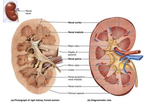
The kidney has three main regions: cortex, medulla, and pelvis.
Renal cortex: Outer, granular region.
Renal medulla: Contains renal pyramids and columns.
Renal pelvis: Funnel-shaped tube continuous with the ureter, collecting urine from calyces.
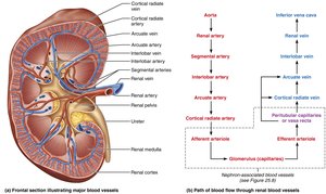
Blood and Nerve Supply
The kidneys receive about 25% of cardiac output. Blood flows through a series of arteries and veins, with the renal artery entering and the renal vein exiting at the hilum. The nerve supply is primarily sympathetic, regulating blood flow and urine formation.
Nephrons: The Functional Units of the Kidney
Structure of Nephrons
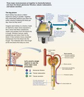
Each kidney contains about one million nephrons, which filter blood and form urine. Each nephron consists of a renal corpuscle and a renal tubule.
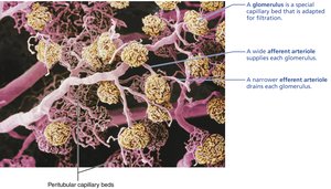
Renal corpuscle: Includes the glomerulus (a tuft of capillaries) and the glomerular (Bowman's) capsule.
Renal tubule: Composed of the proximal convoluted tubule (PCT), nephron loop (loop of Henle), and distal convoluted tubule (DCT).
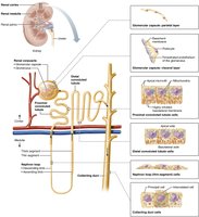
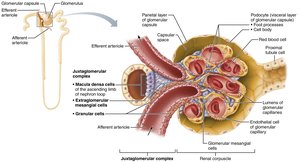
Renal Corpuscle
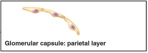
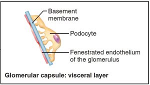
The renal corpuscle is the site of blood filtration. The glomerulus is highly porous, allowing efficient filtrate formation. The glomerular capsule has a parietal layer (simple squamous epithelium) and a visceral layer (podocytes with filtration slits).
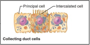
Renal Tubule and Collecting Duct
The renal tubule processes filtrate through reabsorption and secretion. The collecting duct receives filtrate from multiple nephrons and has principal cells (water/Na+ balance) and intercalated cells (acid-base balance).
Classes of Nephrons
Cortical nephrons: 85% of nephrons, mostly in the cortex, with short loops.
Juxtamedullary nephrons: Located near the cortex-medulla junction, with long loops essential for concentrating urine.
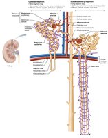
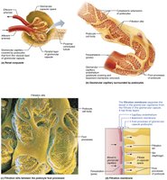
Nephron Capillary Beds
Each nephron is associated with two capillary beds: the glomerulus (for filtration) and the peritubular capillaries or vasa recta (for reabsorption and secretion).
Juxtaglomerular Complex (JGC)
The JGC regulates filtrate formation and blood pressure. It includes macula densa cells (chemoreceptors), granular cells (mechanoreceptors, secrete renin), and extraglomerular mesangial cells (signal transmission).
Urine Formation: Filtration, Reabsorption, and Secretion
Overview of Renal Processes
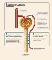
Urine formation involves three main processes:
Glomerular filtration: Passive process where hydrostatic pressure forces fluids and solutes through the filtration membrane.
Tubular reabsorption: Selective movement of substances from filtrate back into blood.
Tubular secretion: Selective movement of substances from blood into filtrate.
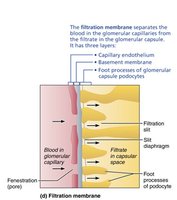
The Filtration Membrane
The filtration membrane consists of three layers: fenestrated endothelium, basement membrane, and podocyte foot processes. It allows passage of water and small solutes but restricts proteins and cells.

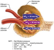
Pressures Affecting Filtration
Filtration is driven by hydrostatic pressure in glomerular capillaries and opposed by hydrostatic pressure in the capsular space and colloid osmotic pressure in capillaries. The net filtration pressure (NFP) determines the glomerular filtration rate (GFR).
Equation:
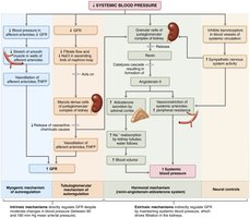
Regulation of Glomerular Filtration Rate (GFR)
GFR is regulated by intrinsic (renal autoregulation) and extrinsic (neural and hormonal) mechanisms. Intrinsic controls include the myogenic mechanism and tubuloglomerular feedback. Extrinsic controls involve the sympathetic nervous system and the renin-angiotensin-aldosterone system.
Tubular Reabsorption and Secretion
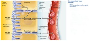
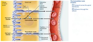
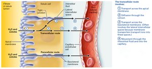
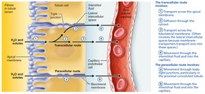
Mechanisms of Tubular Reabsorption
Reabsorption occurs via transcellular (through cells) and paracellular (between cells) routes. It involves both active and passive transport mechanisms.

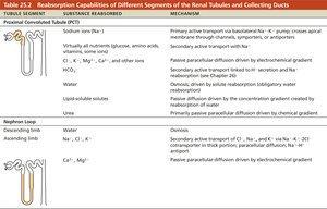
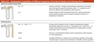
Reabsorptive Capabilities of Renal Tubules and Collecting Ducts
Different segments of the nephron have specialized reabsorptive functions. The PCT is most active, reabsorbing all glucose and amino acids, most water, and ions. The nephron loop and DCT/collecting duct fine-tune reabsorption, regulated by hormones such as ADH, aldosterone, and parathyroid hormone.
Tubular Secretion
Tubular secretion removes additional substances from the blood, including drugs, metabolites, excess potassium, and helps regulate blood pH.
Regulation of Urine Concentration and Volume
Countercurrent Mechanisms
The kidneys use countercurrent mechanisms to regulate urine concentration and volume. The countercurrent multiplier (in nephron loops) creates an osmotic gradient, while the countercurrent exchanger (in vasa recta) preserves it. This allows the kidneys to produce concentrated or dilute urine as needed.
Evaluation of Renal Function
Renal Clearance
Renal clearance is the volume of plasma cleared of a substance per unit time. It is used to assess kidney function and is calculated as:
U: Concentration in urine (mg/mL)
V: Urine flow rate (mL/min)
P: Concentration in plasma (mg/mL)
Urinary Tract Organs: Ureters, Bladder, and Urethra
Ureters
Ureters are muscular tubes that transport urine from the kidneys to the bladder. They have three layers: mucosa, muscularis, and adventitia.
Urinary Bladder
The bladder is a muscular sac for temporary urine storage. It has a mucosa, a thick detrusor muscle, and an adventitia. The trigone is a clinically important region prone to infection.
Urethra
The urethra is a muscular tube that conveys urine out of the body. It is shorter in females and longer in males, with distinct regions in males (prostatic, membranous, and spongy urethra).
Micturition (Urination)
Control of Micturition
Micturition involves the coordinated contraction of the detrusor muscle and relaxation of the internal and external urethral sphincters. It is regulated by autonomic and somatic nervous systems, with voluntary control developing in early childhood.
Developmental Aspects and Clinical Considerations
Development of the Urinary System
The urinary system develops from intermediate mesoderm, with three sets of embryonic kidneys forming in succession. The metanephros becomes the adult kidney.
Clinical Conditions
Pyelonephritis: Kidney infection, often from ascending urinary tract infections.
Renal calculi: Kidney stones, can obstruct urine flow and cause pain.
Urinary tract infections (UTIs): Common, especially in females due to short urethra.
Chronic renal disease and renal failure: Progressive loss of kidney function, requiring dialysis or transplant.
Summary Table: Reabsorption Capabilities of Renal Tubules and Collecting Ducts
Tubule Segment | Substance Reabsorbed | Mechanism |
|---|---|---|
Proximal Convoluted Tubule (PCT) | Na+, all nutrients, water, ions | Primary/secondary active transport, osmosis, passive diffusion |
Nephron Loop | Descending: water Ascending: Na+, Cl-, K+ | Osmosis (descending), active/passive transport (ascending) |
Distal Convoluted Tubule (DCT) | Na+, Cl-, Ca2+ | Active transport, hormone-regulated |
Collecting Duct | Na+, K+, HCO3-, Cl-, water, urea | Active/passive transport, hormone-regulated, facilitated diffusion |
