 Back
The Urinary System: Structure, Function, and Clinical Relevance
Study Guide - Smart Notes
 Tailored notes based on your materials, expanded with key definitions, examples, and context.
The Urinary System
Overview and Functions
The urinary system is essential for maintaining the chemical consistency of blood, filtering waste products, and regulating fluid and electrolyte balance. The kidneys are the primary organs responsible for these functions, filtering blood to remove toxins, metabolic wastes, and excess water, which are then excreted as urine.
Main waste products: Urea, uric acid, and creatinine (all nitrogenous wastes).
Key functions of the kidneys:
Regulate blood volume and pressure
Control electrolyte and acid-base balance
Remove metabolic wastes
Organs of the Urinary System
Kidneys
Ureters
Urinary bladder
Urethra
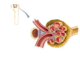
Location and External Anatomy of the Kidneys
Position and Structure
The kidneys are retroperitoneal organs, located behind the peritoneum, lateral to the T12–L3 vertebrae. Each kidney is approximately 12 cm tall, 6 cm wide, and 3 cm thick. The hilum is the entry and exit site for vessels, nerves, and the ureter.
Color: Red-brown
Protective layers:
Fibrous capsule (dense connective tissue)
Perirenal fat capsule (external to capsule)
Renal fascia (external to fat capsule, contains pararenal fat)
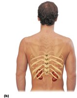
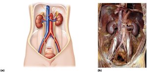
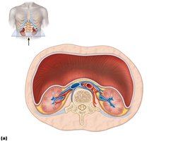
Internal Gross Anatomy of the Kidneys
Regions and Structures
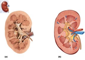
A frontal section of the kidney reveals two main regions: the renal cortex (outer, granular) and the renal medulla (inner, composed of cone-shaped pyramids). The renal pelvis collects urine from major and minor calices and channels it to the ureter.
Renal cortex: Superficial, granular appearance
Renal medulla: Contains renal pyramids
Renal pelvis: Funnel-shaped tube continuous with ureter
Major and minor calices: Collect urine from pyramids
Renal Vasculature
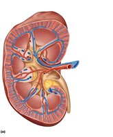
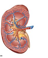
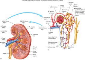
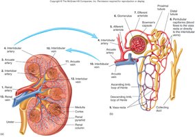
The kidneys receive blood from the renal arteries, which branch into segmental, interlobar, arcuate, and cortical radiate arteries. Blood is filtered in the glomerulus and returns via corresponding veins.
Renal artery → Segmental artery → Interlobar artery → Arcuate artery → Cortical radiate artery
Renal vein returns filtered blood to the inferior vena cava
Microscopic Anatomy of the Kidneys
The Nephron
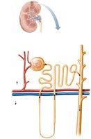
The nephron is the functional unit of the kidney, with over one million per kidney. Each nephron consists of a renal corpuscle and a renal tubule.
Renal corpuscle: Site of blood filtration (glomerulus + glomerular capsule)
Renal tubule: Processes filtrate into urine
Mechanisms of Urine Production
Filtration: Blood plasma is filtered from glomerular capillaries into the nephron
Resorption: Essential nutrients, water, and ions are reclaimed
Secretion: Additional wastes are actively transported into the filtrate
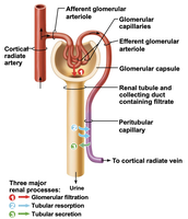
Renal Corpuscle and Filtration Membrane
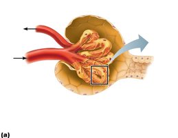
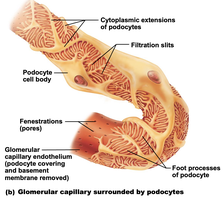
The renal corpuscle consists of the glomerulus (a tuft of fenestrated capillaries) and the glomerular (Bowman's) capsule. The filtration membrane allows passage of water, ions, glucose, amino acids, and urea, but holds back most proteins.
Three layers of the filtration membrane:
Fenestrated endothelium
Basement membrane
Filtration slits between podocyte foot processes
Renal Tubule and Collecting Ducts
Filtrate passes from the glomerulus into the renal tubule, which consists of the proximal convoluted tubule, nephron loop (descending and ascending limbs), distal convoluted tubule, and collecting duct. Collecting ducts play a key role in water conservation, regulated by antidiuretic hormone (ADH).
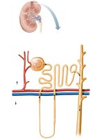
Types of Nephrons
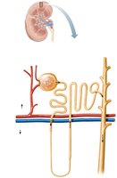
Cortical nephrons: Short nephron loops, glomerulus further from cortex-medulla junction, efferent arteriole supplies peritubular capillaries
Juxtamedullary nephrons: Long nephron loops, glomerulus closer to cortex-medulla junction, efferent arteriole supplies vasa recta
Blood Vessels Associated with Nephrons
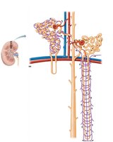
Glomerulus: Produces filtrate; fed and drained by afferent and efferent arterioles
Peritubular capillaries: Adapted for absorption, surround cortical nephrons
Vasa recta: Long, looping vessels associated with juxtamedullary nephrons, crucial for urine concentration
Juxtaglomerular Complex
Structure and Function
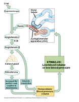
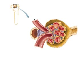
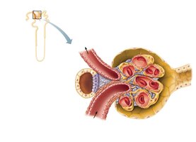
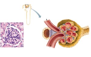
The juxtaglomerular complex regulates blood pressure and filtrate formation. It consists of granular cells (secrete renin), macula densa (monitor sodium concentration), and mesangial cells (regulate blood flow).
Granular cells: Release renin in response to low blood pressure
Macula densa: Signal granular cells to secrete renin
Mesangial cells: Regulate glomerular blood flow
Ureters
Structure and Function
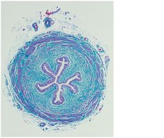
Ureters are muscular tubes that transport urine from the kidneys to the urinary bladder. Their oblique entry into the bladder prevents backflow of urine.
Mucosa: Transitional epithelium
Muscularis: Inner longitudinal and outer circular smooth muscle layers
Adventitia: Connective tissue
Urinary Bladder
Structure and Function
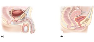
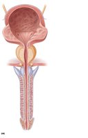
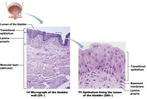
The urinary bladder is a collapsible muscular sac that stores and expels urine. When full, it expands into the abdominal cavity; when empty, it lies within the pelvis. In males, the prostate gland lies inferior to the bladder and surrounds the urethra.
Layers:
Mucosa (transitional epithelium)
Thick muscular layer (detrusor muscle)
Adventitia
Trigone: Triangular area between ureteric orifices and urethra
Urethra
Structure and Function
The urethra conveys urine from the bladder to the exterior. Its epithelium transitions from transitional (proximal) to stratified squamous (distal). The internal urethral sphincter is involuntary smooth muscle, while the external sphincter is voluntary skeletal muscle.
Female urethra: 3–4 cm long
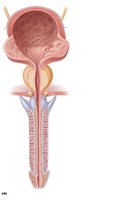
Male urethra: 20 cm long, with prostatic, intermediate, and spongy regions
Micturition (Urination)
Micturition is the act of emptying the bladder, controlled by both autonomic and somatic nervous systems. The process involves the relaxation of the internal and external urethral sphincters and contraction of the detrusor muscle.
Disorders of the Urinary System
Urinary tract infections (UTIs): More common in females; symptoms include burning during urination
Renal calculi: Kidney stones
Bladder cancer: More common in men, accounts for 3% of cancers
Kidney cancer: Originates from epithelial cells of uriniferous tubules
