 Back
BackThe Urinary System: Structure, Function, and Clinical Relevance
Study Guide - Smart Notes
 Tailored notes based on your materials, expanded with key definitions, examples, and context.
The Urinary System: Overview and Major Functions
Introduction to the Urinary System
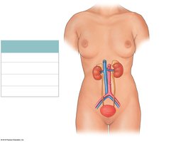
The urinary system is essential for maintaining homeostasis by regulating the composition and volume of blood, removing metabolic wastes, and balancing fluids and electrolytes. It consists of the kidneys, ureters, urinary bladder, and urethra.
Kidneys: Filter blood, remove wastes, and produce urine.
Ureters: Transport urine from the kidneys to the bladder.
Urinary bladder: Stores urine until elimination.
Urethra: Conducts urine from the bladder to the exterior.
Major Functions of the Urinary System
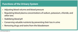
Adjusting blood volume and blood pressure: By regulating water excretion and hormone release.
Regulating plasma ionic composition: Controls levels of sodium, potassium, chloride, and other ions.
Stabilizing blood pH: By excreting hydrogen ions and reabsorbing bicarbonate.
Conserving valuable nutrients: Prevents loss of glucose, amino acids, and other essential molecules.
Removing drugs and toxins: Eliminates metabolic wastes and foreign substances from the bloodstream.
Kidney Anatomy and Position
Location and Structural Features of the Kidneys
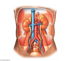
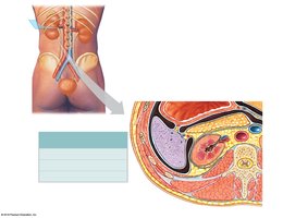
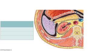
The kidneys are retroperitoneal organs located on either side of the vertebral column, between the T12 and L3 vertebrae. The left kidney is slightly superior to the right. Each kidney is protected by the 11th and 12th ribs and surrounded by connective tissue layers for support and protection.
Hilum: Medial indentation for entry/exit of renal artery, vein, and ureter.
Connective tissue layers: Fibrous capsule (innermost), perinephric fat, and renal fascia (outermost).
Gross Structure of the Kidney
Internal Anatomy
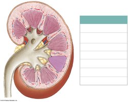
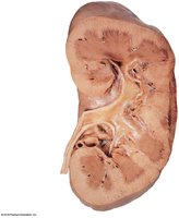
Internally, the kidney consists of the renal cortex, renal medulla (containing pyramids and papillae), renal columns, calyces, and renal pelvis. The cortex and medulla form the functional tissue (parenchyma) of the kidney.
Renal cortex: Outer region containing most nephrons.
Renal medulla: Inner region with renal pyramids and papillae.
Renal pelvis: Funnel-shaped structure collecting urine from calyces.
Nephrons: Structure and Function
Types of Nephrons
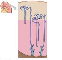
Nephrons are the microscopic functional units of the kidney. There are two main types:
Cortical nephrons (85%): Located mostly in the cortex; responsible for most regulatory functions.
Juxtamedullary nephrons (15%): Have long loops extending deep into the medulla; crucial for establishing the osmotic gradient necessary for urine concentration.
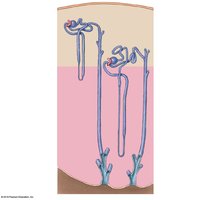
Regions of the Nephron and Collecting System
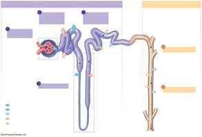
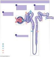
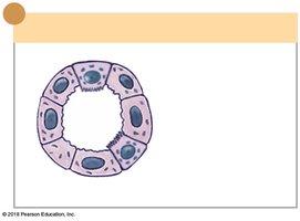
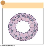
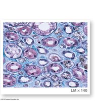
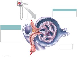
Each nephron consists of a renal corpuscle and a renal tubule. The renal corpuscle includes the glomerulus and glomerular capsule, where filtration occurs. The renal tubule is divided into the proximal convoluted tubule (PCT), nephron loop (loop of Henle), and distal convoluted tubule (DCT). The collecting system receives fluid from multiple nephrons and delivers urine to the renal pelvis.
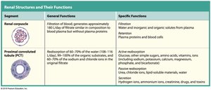
Renal corpuscle: Site of filtration; produces protein-free filtrate.
PCT: Reabsorbs nutrients, ions, and water from filtrate.
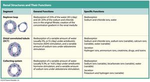
Nephron loop: Establishes osmotic gradient in the medulla.
DCT: Further adjusts filtrate by secretion and reabsorption.
Collecting duct: Final adjustments to urine composition and volume.
Blood Flow in the Kidney
Pathway of Blood Flow
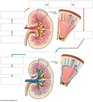
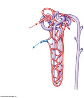
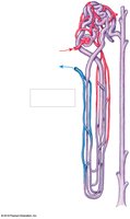
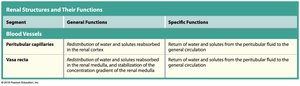
Blood enters the kidney via the renal artery, passes through segmental, interlobar, arcuate, and cortical radiate arteries, and then into afferent arterioles. Filtration occurs in the glomerulus, and blood exits via efferent arterioles. In cortical nephrons, efferent arterioles form peritubular capillaries; in juxtamedullary nephrons, they form the vasa recta, which are essential for maintaining the medullary osmotic gradient.
Renal Physiology: Filtration, Reabsorption, and Secretion
Overview of Renal Processes
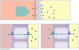
The kidneys maintain homeostasis by filtering blood, reabsorbing essential substances, and secreting wastes. The three main processes are:
Filtration: Blood pressure forces water and solutes from glomerular capillaries into the nephron.
Reabsorption: Movement of water and solutes from the nephron back into the blood.
Secretion: Transfer of additional wastes from blood into the nephron for excretion.
Major Functions of Nephron Segments
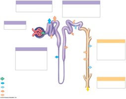
Each segment of the nephron and collecting system has specialized functions in filtration, reabsorption, and secretion.
Segment | General Functions | Specific Functions |
|---|---|---|
Renal corpuscle | Filtration of blood; generates filtrate | Retention of plasma proteins and blood cells |
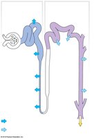
Proximal convoluted tubule (PCT) | Reabsorption of most water, ions, and nutrients | Active reabsorption of glucose, amino acids, vitamins, ions; secretion of H+, ammonia, creatinine, drugs |
Nephron loop | Reabsorption of water and ions; creation of medullary gradient | Reabsorption of sodium, chloride, water |
Distal convoluted tubule (DCT) | Variable reabsorption and secretion; hormonal regulation | Reabsorption of sodium, calcium, water; secretion of H+, ammonia, drugs |
Collecting system | Final adjustments of urine; variable water reabsorption | Reabsorption of water, sodium, bicarbonate; secretion of potassium, hydrogen |
Filtration Membrane and Filtration Pressure
Structure of the Renal Corpuscle and Filtration Membrane
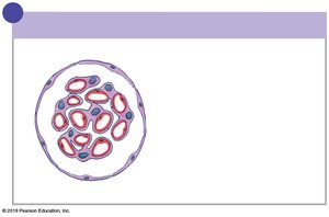
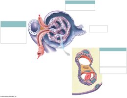
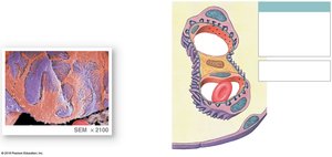
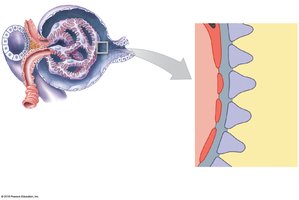
The renal corpuscle consists of the glomerulus and glomerular capsule. The filtration membrane is formed by the fenestrated endothelium of glomerular capillaries, a basement membrane, and the foot processes of podocytes. This structure allows passage of water and small solutes but restricts proteins and cells.
Filtration Pressure
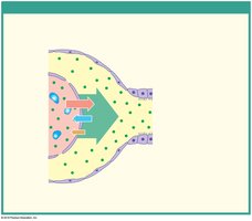
Filtration pressure (FP) is the net force driving filtration at the glomerulus. It is determined by:
Glomerular hydrostatic pressure (GHP): Pushes water and solutes out of plasma.
Blood colloid osmotic pressure (BCOP): Draws water back into plasma.
Capsular hydrostatic pressure (CsHP): Opposes filtration by pushing water back into plasma.
The equation for net filtration pressure is:
Regulation of Glomerular Filtration Rate (GFR)
Autoregulation and Central Regulation
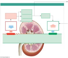
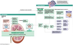
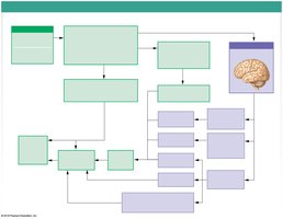
The glomerular filtration rate (GFR) is tightly regulated by local (autoregulation) and systemic (central) mechanisms.
Autoregulation: Adjusts afferent/efferent arteriole diameter and glomerular capillary surface area in response to changes in blood flow or pressure.
Central regulation: Involves the renin-angiotensin-aldosterone system (RAAS) and sympathetic nervous system to restore GFR if autoregulation fails.
Transport Mechanisms in the Nephron
Proximal Convoluted Tubule (PCT)
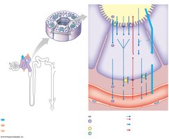
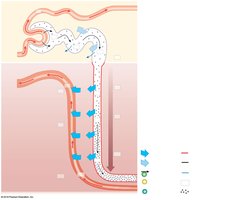
The PCT is responsible for the majority of solute and water reabsorption. Sodium transporters facilitate the reabsorption of glucose, amino acids, bicarbonate, and other ions, with water following by osmosis.
Distal Convoluted Tubule (DCT)
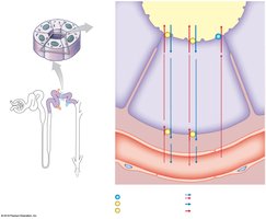
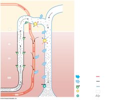
The DCT fine-tunes the filtrate by reabsorbing sodium (in exchange for potassium, regulated by aldosterone) and secreting hydrogen ions, drugs, and toxins.
Countercurrent Multiplication and Urine Concentration
Countercurrent Multiplication in the Nephron Loop
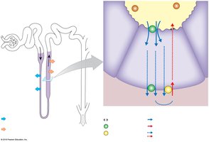
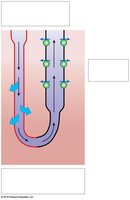
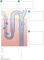
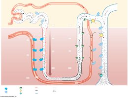
Countercurrent multiplication in the nephron loop establishes the osmotic gradient in the renal medulla, which is essential for concentrating urine. The descending limb is permeable to water but not solutes, while the ascending limb is permeable to solutes but not water. This arrangement allows for efficient reabsorption of water and solutes.
Hormonal Regulation of Urine Volume and Concentration
Role of Antidiuretic Hormone (ADH)
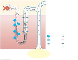
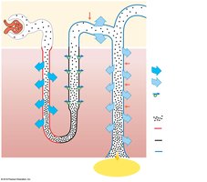
ADH regulates facultative water reabsorption in the DCT and collecting duct. In the presence of ADH, aquaporins are inserted into the membranes, increasing water reabsorption and producing concentrated urine. Without ADH, the urine is dilute.
Summary of Water Reabsorption and Urine Production
Filtrate is modified as it passes through the nephron and collecting system. The final urine concentration depends on the actions of the nephron segments, collecting ducts, and the presence of hormones such as ADH and aldosterone. The vasa recta help maintain the medullary osmotic gradient.
Clinical Considerations: Renal Failure and Dialysis
Types of Renal Failure
Acute renal failure: Sudden loss of kidney function, often reversible if treated promptly.
Chronic renal failure: Gradual loss of function, usually irreversible; managed by dietary restrictions and, in severe cases, dialysis or transplantation.
Hemodialysis
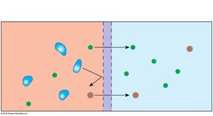
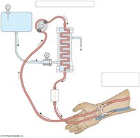
Hemodialysis is a process where blood is filtered through an artificial membrane to remove wastes and excess substances. It is used in patients with severe renal failure.
Urine Transport, Storage, and Elimination
Urinary Tract Structures
Ureters: Muscular tubes transporting urine from kidneys to bladder by peristalsis.
Urinary bladder: Hollow organ for temporary urine storage; contains detrusor muscle for contraction.
Urethra: Tube conducting urine to the exterior; longer in males and also transports semen.
Histology of the Urinary Tract
The ureters and bladder are lined with transitional epithelium, allowing for expansion. The bladder wall contains smooth muscle (detrusor muscle), and the urethra is lined with stratified epithelium.
Control of Urination (Micturition Reflex)
Neural Control
The micturition reflex involves both local (spinal) and central (cerebral cortex) pathways. Stretch receptors in the bladder wall trigger reflex contraction of the detrusor muscle and relaxation of the internal and external urethral sphincters, allowing urination when appropriate.
Common Urinary Disorders
Symptoms and Conditions
Pain: Flank pain may indicate kidney infection or stones; suprapubic pain may indicate bladder disorders.
Dysuria: Painful urination, often due to infection or obstruction.
Polyuria: Excessive urine output (e.g., diabetes).
Oliguria: Low urine output (50–500 mL/day).
Anuria: Very low or absent urine output (<50 mL/day).
Incontinence: Inability to control urination.
Urinary retention: Inability to void despite normal kidney function.
Medical Terminology
Azotemia: Presence of nitrogenous wastes in the blood.
Hydronephrosis: Swelling of the kidney due to urine outflow obstruction.
Uremia: Toxic accumulation of urea in the blood due to kidney failure.
