Back
BackThe Urinary System: Structure, Function, and Physiology
Study Guide - Smart Notes
 Tailored notes based on your materials, expanded with key definitions, examples, and context.
The Urinary System
Introduction
The urinary system is essential for maintaining the body's internal environment by regulating the volume and composition of blood, removing metabolic wastes, and balancing electrolytes and pH. It consists of several organs that work together to produce, transport, store, and eliminate urine.
Functions: Excretion of metabolic wastes, regulation of blood volume and pressure, regulation of blood pH, and maintenance of electrolyte balance.
Organs: Kidneys, ureters, urinary bladder, and urethra.
Kidney Anatomy
Location and External Anatomy
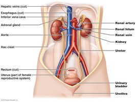
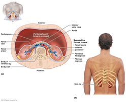
The kidneys are retroperitoneal organs located on either side of the vertebral column, with the right kidney slightly lower due to the position of the liver. They are protected by the lower ribs and surrounded by supportive tissue layers.
Supportive Layers: Fibrous capsule, perirenal fat capsule, and renal fascia.
Renal Hilum: The entry and exit site for the renal artery, vein, and ureter.
Internal Anatomy
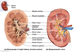
The kidney is divided into three main regions: the cortex, medulla, and pelvis. The cortex is the outer region, the medulla contains renal pyramids, and the pelvis collects urine before it enters the ureter.
Renal Cortex: Outer region containing renal corpuscles and convoluted tubules.
Renal Medulla: Inner region with renal pyramids and columns.
Renal Pelvis: Funnel-shaped structure that collects urine and channels it into the ureter.
Blood and Nerve Supply
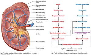
The kidneys receive about 25% of the cardiac output through the renal arteries. Blood is filtered in the nephrons and returned via the renal veins. The renal plexus provides nerve supply, primarily sympathetic fibers.
Renal Arteries: Branch from the aorta and supply blood to the kidneys.
Renal Veins: Drain filtered blood into the inferior vena cava.
Nephrons: The Functional Unit of the Kidney
Structure of the Nephron
Each kidney contains about 1 million nephrons, which are responsible for filtering blood and forming urine. Nephrons consist of a renal corpuscle and a renal tubule.
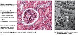
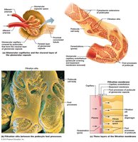
Renal Corpuscle: Includes the glomerulus (a tuft of capillaries) and Bowman's (glomerular) capsule.
Renal Tubule: Composed of the proximal convoluted tubule (PCT), loop of Henle (nephron loop), distal convoluted tubule (DCT), and collecting duct.
Types of Nephrons
Cortical Nephrons: Located mostly in the cortex, with short loops of Henle.
Juxtamedullary Nephrons: Located near the cortex-medulla junction, with long loops of Henle that extend deep into the medulla, crucial for concentrating urine.
Renal Corpuscle and Filtration Membrane
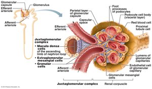
The renal corpuscle is the site of blood filtration. The filtration membrane consists of three layers: fenestrated endothelium, basement membrane, and podocyte foot processes. This structure allows water and small solutes to pass while retaining blood cells and large proteins.
Juxtaglomerular Apparatus
This specialized structure regulates blood pressure and filtration rate. It includes juxtaglomerular (granular) cells, macula densa cells, and extraglomerular mesangial cells.
Juxtaglomerular Cells: Secrete renin in response to low blood pressure.
Macula Densa: Detects sodium concentration in the distal tubule.
Kidney Physiology: Mechanism of Urine Formation
Three Major Renal Processes
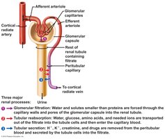
Glomerular Filtration: Blood pressure forces water and solutes from the glomerulus into the glomerular capsule, forming filtrate.
Tubular Reabsorption: Useful substances are reclaimed from the filtrate back into the blood, mainly in the PCT.
Tubular Secretion: Additional wastes and excess ions are secreted from the blood into the filtrate.
Glomerular Filtration and Net Filtration Pressure (NFP)
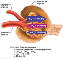
Filtration at the glomerulus depends on the balance of hydrostatic and osmotic pressures. The net filtration pressure (NFP) determines the amount of filtrate formed.
Glomerular Hydrostatic Pressure (HPgc): Pushes water and solutes out of the blood (about 55 mm Hg).
Blood Colloid Osmotic Pressure (OPgc): Pulls water back into the blood (about 30 mm Hg).
Capsular Hydrostatic Pressure (HPcs): Opposes filtration (about 15 mm Hg).
Net Filtration Pressure:
Glomerular Filtration Rate (GFR) and Its Regulation
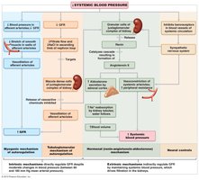
GFR is the volume of filtrate formed per minute by both kidneys. It is tightly regulated by intrinsic (renal autoregulation) and extrinsic (neural and hormonal) mechanisms to ensure homeostasis.
Intrinsic Controls: Myogenic mechanism and tubuloglomerular feedback maintain GFR despite moderate changes in blood pressure.
Extrinsic Controls: Sympathetic nervous system and renin-angiotensin-aldosterone system (RAAS) regulate GFR during extreme conditions.
Tubular Reabsorption and Secretion
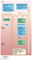
Most filtrate is reabsorbed in the renal tubules, with different segments specialized for reabsorbing specific substances. Tubular secretion removes additional wastes and helps regulate blood pH.
Proximal Convoluted Tubule (PCT): Reabsorbs 65% of filtrate, including water, sodium, glucose, and amino acids.
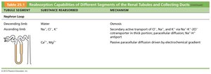
Loop of Henle: Descending limb reabsorbs water; ascending limb reabsorbs NaCl but is impermeable to water.
Distal Convoluted Tubule (DCT) and Collecting Duct: Fine-tune reabsorption under hormonal control (aldosterone, ADH).
Countercurrent Mechanism and Medullary Osmotic Gradient
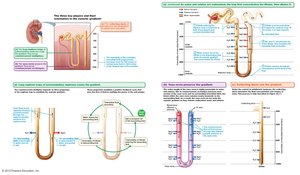
The countercurrent multiplier (in the loop of Henle) and countercurrent exchanger (in the vasa recta) establish and maintain a medullary osmotic gradient, allowing the kidneys to concentrate or dilute urine as needed.
Descending Limb: Permeable to water, not solutes; water leaves, concentrating filtrate.
Ascending Limb: Impermeable to water, actively transports NaCl out; dilutes filtrate.
Vasa Recta: Maintains the gradient by exchanging water and solutes with the interstitial fluid.
Regulation of Urine Concentration and Volume
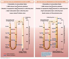
Urine concentration is regulated by antidiuretic hormone (ADH) and aldosterone. ADH increases water reabsorption in the collecting ducts, producing concentrated urine. In the absence of ADH, urine is dilute.
Concentrated Urine: High ADH, water reabsorbed, urine up to 1200 mOsm.
Dilute Urine: Low ADH, little water reabsorbed, urine as low as 100 mOsm.
Renal Clearance
Renal clearance is the volume of plasma cleared of a substance per unit time. It is used to assess kidney function. Inulin is used as a standard because it is freely filtered, not reabsorbed, and not secreted.
Clearance greater than inulin: Substance is secreted.
Clearance less than inulin: Substance is reabsorbed.
Characteristics and Composition of Urine
Physical Characteristics
Color: Ranges from pale yellow to deep amber depending on concentration.
Odor: Slightly aromatic; can be affected by diet or disease.
pH: Usually around 6, but ranges from 4.5 to 8.
Specific Gravity: 1.001–1.035 (denser than water).
Chemical Composition
Water: About 95% of urine volume.
Solutes: Urea, creatinine, uric acid, ions (Na+, K+, Cl-, etc.).
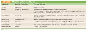
Abnormal Urinary Constituents
Substance | Name of Condition | Possible Causes |
|---|---|---|
Glucose | Glycosuria | Diabetes mellitus |
Proteins | Proteinuria, albuminuria | Excessive exertion, pregnancy, glomerulonephritis, hypertension, renal disease |
Ketone bodies | Ketonuria | Starvation, untreated diabetes mellitus |
Hemoglobin | Hemoglobinuria | Transfusion reaction, hemolytic anemia, burns |
Bile pigments | Bilirubinuria | Liver disease, bile duct obstruction |
Erythrocytes | Hematuria | Trauma, kidney stones, infection, cancer |
Leukocytes (pus) | Pyuria | Urinary tract infection |
Ureters
Histology
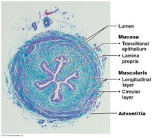
The ureter wall consists of three layers: mucosa (transitional epithelium), muscularis (smooth muscle), and adventitia (connective tissue). These layers facilitate the transport of urine from the kidneys to the bladder by peristalsis and gravity.
Urinary Bladder
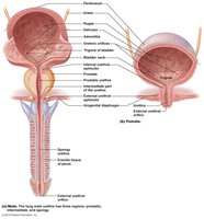
The urinary bladder is a muscular sac that stores urine until micturition (voiding). Its wall contains rugae for expansion and the detrusor muscle for contraction. The trigone is a triangular area defined by the openings of the ureters and urethra.
Urethra
The urethra conveys urine from the bladder to the outside of the body. It has internal (smooth muscle, involuntary) and external (skeletal muscle, voluntary) sphincters. The male urethra is longer and divided into prostatic, membranous, and spongy regions; the female urethra is shorter.
Micturition (Urination)
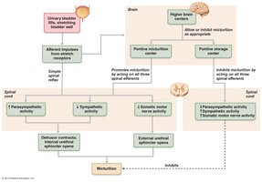
Micturition is the process of voiding urine. It is controlled by a spinal reflex, with voluntary control from higher brain centers. Stretch receptors in the bladder wall trigger the reflex when the bladder fills to about 200 mL.
Parasympathetic activity: Contracts detrusor muscle, opens internal sphincter.
Somatic motor activity: Relaxes external sphincter for urination.
Higher brain centers: Can inhibit or facilitate micturition.
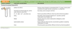
Summary Table: Reabsorption Capabilities of Renal Tubules
Tubule Segment | Substance Reabsorbed | Mechanism |
|---|---|---|
Proximal Convoluted Tubule (PCT) | Na+, nutrients, Cl-, K+, HCO3-, water, lipid-soluble solutes, urea | Primary active transport, passive paracellular diffusion, osmosis, secondary active transport, passive diffusion |
Nephron Loop (Descending limb) | Water | Osmosis |
Nephron Loop (Ascending limb) | Na+, Cl-, K+ | Secondary active transport, passive paracellular diffusion |
Distal Convoluted Tubule (DCT) and Collecting Duct | Na+, Cl-, Ca2+, water, urea | Regulated by hormones (aldosterone, ADH, PTH), osmosis, passive diffusion |