 Back
The Urinary System: Structure, Function, and Regulation
Study Guide - Smart Notes
 Tailored notes based on your materials, expanded with key definitions, examples, and context.
The Urinary System
Components and Functions of the Urinary System
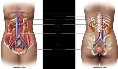
The urinary system is essential for maintaining homeostasis by filtering blood, removing wastes, and regulating fluid and electrolyte balance. It consists of several organs working together to produce, store, and eliminate urine.
Kidneys: Filter blood and produce urine.
Ureters: Transport urine from kidneys to bladder.
Urinary bladder: Stores urine until excretion.
Urethra: Conducts urine out of the body.
Key Functions:
Excretion of toxic metabolic wastes
Regulation of blood volume, pressure, and osmolarity
Electrolyte and acid-base balance
Secretion of erythropoietin (stimulates red blood cell production)
Final step in synthesizing calcitriol (active vitamin D)
Detoxification of some drugs
Excretion and Wastes
Excretion is the process of separating and eliminating wastes from body fluids.
Waste: Any substance that is useless or in excess of the body’s needs.
Metabolic waste: Produced by the body’s metabolic processes.
Nitrogenous wastes: Highly toxic; includes urea (50%), uric acid, and creatinine.
Nephrons: The Functional Unit of the Kidney
Structure of the Nephron
Nephrons are microscopic structures responsible for urine formation. Each nephron consists of two main parts:
Renal corpuscle: Site of blood filtration.
Renal tubule: Processes filtrate into urine.
Renal Corpuscle
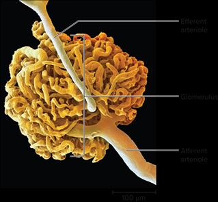
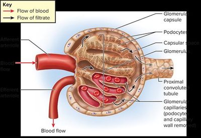
The renal corpuscle is composed of a ball of capillaries called the glomerulus, surrounded by a two-layered glomerular capsule.
Capsular space: Separates the capsule layers.
Afferent arteriole: Brings blood into the glomerulus (larger diameter).
Efferent arteriole: Takes blood away (smaller diameter, creates high pressure).
Renal Tubule
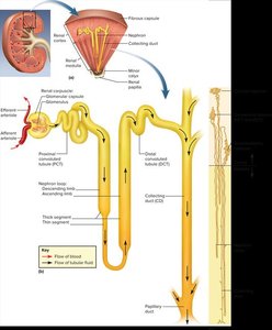
The renal tubule is divided into four regions, each with unique properties and roles in urine production:
Proximal convoluted tubule (PCT): Absorbs nutrients like glucose, amino acids, and small peptides; lined with cuboidal epithelium and microvilli.
Nephron loop (Loop of Henle): U-shaped; descending limb (permeable to water), ascending limb (active ion transport).
Distal convoluted tubule (DCT): Responsible for sodium and chloride reabsorption.
Collecting duct (CD): Receives fluid from multiple nephrons; mainly reabsorbs water; urine is officially formed here.
Urine Formation: Processes and Regulation
Stages of Urine Formation
The kidneys convert blood plasma to urine in four stages:
Glomerular filtration: Water and solutes are filtered from blood into the nephron.
Tubular reabsorption: Useful substances are reabsorbed into the blood.
Tubular secretion: Additional wastes are secreted into the tubule.
Water conservation: Water is reabsorbed to concentrate urine.
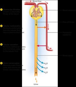
Glomerular Filtration
Glomerular filtration is the first step in urine formation. The filtration membrane consists of:
Endothelium of capillary: Fenestrated, allows passage of small molecules but not blood cells.
Basement membrane: Glycoprotein gel, blocks large proteins.
Filtration slits: Formed by podocytes, restrict passage of large molecules.
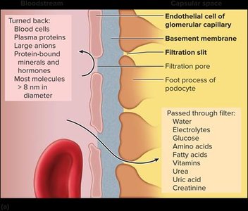
Filtration Pressure
Filtration is driven by blood pressure, which is higher in the glomerulus than in other capillaries.
High pressure: Forces fluid into the capsular space.
Kidney vulnerability: Hypertension can damage glomerular capillaries, leading to renal failure.
Glomerular Filtration Rate (GFR)
Definition: Amount of filtrate formed per minute by both kidneys.
Normal values: Females ~105 mL/min (150L/day), Males ~125 mL/min (180L/day).
Urine output: Only 1-2L/day; 99% of filtrate is reabsorbed.
Regulation of GFR
GFR must be tightly regulated to maintain homeostasis.
Too high: Dehydration and electrolyte depletion.
Too low: Wastes are reabsorbed instead of eliminated.
Regulation mechanisms:
Neural control (sympathetic constriction of afferent arterioles)
Hormonal control (renin-angiotensin-aldosterone system)
Local control (renal autoregulation)
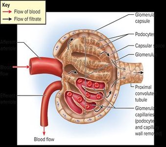
Renal Autoregulation
Nephrons can independently regulate their own GFR via the juxtaglomerular apparatus, which monitors flow rate and osmolarity.
Macula densa: Sensory cells in the DCT.
Granular cells: Secrete renin in response to changes in blood pressure.
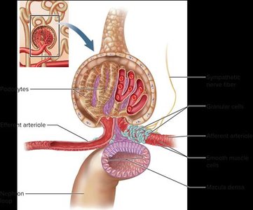
Summary Table: Components and Functions of the Urinary System
Component | Main Function |
|---|---|
Kidneys | Filter blood, produce urine, regulate fluid/electrolyte balance |
Ureters | Transport urine to bladder |
Urinary bladder | Store urine |
Urethra | Conduct urine out of body |
Key Terms and Concepts
Nephron: Functional unit of the kidney
Glomerulus: Capillary network for filtration
Glomerular filtration rate (GFR): Rate of filtrate formation
Renin-angiotensin-aldosterone system: Hormonal regulation of blood pressure and GFR
Juxtaglomerular apparatus: Local regulation of GFR
Relevant Equations
GFR Calculation:
Net Filtration Pressure:
Example: Clinical Application
Hypertension: Chronic high blood pressure can damage glomerular capillaries, leading to renal failure.
Diabetes: Excess glucose can overwhelm reabsorption in the PCT, leading to glucose in urine (glycosuria).
Additional info:
Renal autoregulation involves feedback mechanisms that adjust afferent and efferent arteriole diameter to stabilize GFR.
Hormonal regulation via aldosterone and ADH helps conserve water and maintain blood pressure.
