 Back
BackThe Urinary System: Structure, Function, and Clinical Relevance
Study Guide - Smart Notes
 Tailored notes based on your materials, expanded with key definitions, examples, and context.
Urinary System Overview
Introduction
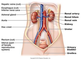
The urinary system is essential for filtering blood, removing waste products, and maintaining homeostasis of water, electrolytes, and acid-base balance. It consists of the kidneys, ureters, urinary bladder, and urethra.
Kidney Anatomy and Location
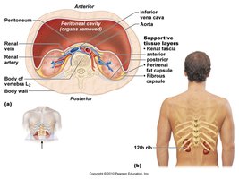
Location and Protective Structures
Location: Retroperitoneal, spanning from T12 to L3 vertebrae.
Size: Approximately the size of a bar of soap.
Protection: Surrounded by an adipose capsule and fibrous capsule, which protect and anchor the kidneys.
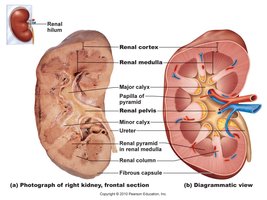
Gross Anatomy of the Kidney
Renal Cortex: The outer region of the kidney.
Renal Medulla: The inner region, containing renal pyramids.
Renal Pyramid: Cone-shaped tissues in the medulla.
Renal Column: Extensions of cortex between pyramids.
Renal Pelvis: Funnel-shaped structure collecting urine into the ureter.
Calyces: Minor and major calyces collect urine from pyramids.
Kidney Functions
Major Functions
Filtration: Removes waste from blood plasma.
Homeostasis: Regulates osmolarity, blood volume, blood pressure, and acid-base balance.
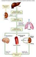
Secretion: Produces hormones such as renin (regulates BP) and erythropoietin (stimulates RBC production).
Detoxification: Metabolizes drugs and free radicals.
Nephron Structure and Function
Nephron Overview
Nephrons are the functional units of the kidney, responsible for urine formation. Each kidney contains about 1.2 million nephrons.
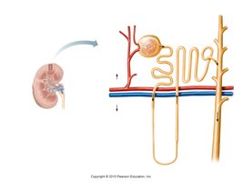
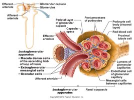
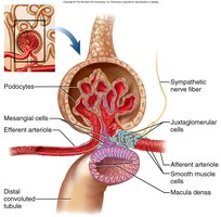
Renal Corpuscle
Components: Glomerulus (capillary bed) and glomerular (Bowman's) capsule.
Filtration: Blood pressure forces small molecules through the filtration membrane; large molecules remain in the blood.
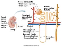
Renal Tubule
Segments: Proximal convoluted tubule (PCT), nephron loop (Loop of Henle), distal convoluted tubule (DCT), and collecting duct (CD).
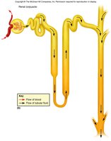
Flow of Filtrate: Glomerular capsule → PCT → nephron loop → DCT → collecting duct → minor calyx → major calyx → renal pelvis → ureter → bladder → urethra.
Types of Nephrons
Cortical Nephrons (85%): Short loops, located near the kidney surface.
Juxtamedullary Nephrons (15%): Long loops extending deep into the medulla, crucial for concentrating urine.
Urine Formation
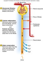
Three Main Processes
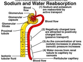
Glomerular Filtration: Movement of fluid and solutes from blood into the nephron.
Tubular Reabsorption: Return of water and solutes from filtrate to blood.
Tubular Secretion: Addition of wastes from blood to filtrate.
Filtration Membrane
Fenestrated Endothelium: Excludes blood cells and large proteins.
Basement Membrane: Proteoglycan gel, blocks large molecules.
Filtration Slits: Podocyte extensions allow only small particles (water, electrolytes, glucose, amino acids, nitrogenous wastes, vitamins) to pass.
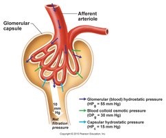
Filtration Pressure
High pressure in the glomerulus (due to wider afferent arteriole) drives filtration.
Filtration pressure overrides reabsorption, ensuring continuous filtration.
Glomerular Filtration Rate (GFR)
Definition: Volume of filtrate formed per minute by both kidneys.
Normal Values: ~125 mL/min (180 L/day) in males; ~105 mL/min (150 L/day) in females.
Clinical Relevance: 99% of filtrate is reabsorbed; only 1–2 L urine excreted daily. High GFR can cause dehydration; low GFR leads to waste retention (anuria).
Regulation of Glomerular Filtration
Autoregulation
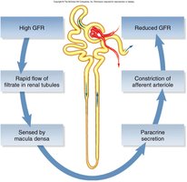
Myogenic Mechanism: Increased BP stretches afferent arteriole, causing constriction to stabilize GFR.
Tubuloglomerular Feedback: Macula densa cells in DCT sense flow and signal juxtaglomerular cells to adjust arteriole diameter.
Sympathetic and Hormonal Control
Sympathetic Control: During stress, afferent arterioles constrict, reducing GFR and urine output to redirect blood to vital organs.
Hormonal Control: Renin-angiotensin-aldosterone system (RAAS) increases BP and blood volume by promoting sodium and water reabsorption.
Tubular Reabsorption and Secretion
Reabsorption in the Proximal Convoluted Tubule (PCT)
Major Site: Most water, nutrients (glucose, amino acids, vitamins), and ions are reabsorbed here.
Mechanisms: Active and passive transport, solvent drag.
Tubular Secretion
Purpose: Removes additional wastes (urea, uric acid, ammonia, drugs) and regulates acid-base balance (H+, bicarbonate).
Direction: Substances move from peritubular capillaries into the nephron tubule.
Nephron Loop (Loop of Henle)
Descending Limb: Permeable to water, not electrolytes.
Ascending Limb: Permeable to electrolytes (Na+, Cl-, K+), not water.
Function: Creates a salinity gradient, allowing the collecting duct to concentrate urine.
DCT and Collecting Duct
Regulation: Hormones (aldosterone, ADH) control reabsorption of water and salts, affecting urine volume and concentration.
Acid-Base Balance: Secretion and reabsorption of H+ and bicarbonate ions.

Summary Table: Tubular Reabsorption and Secretion
Segment | Main Function | Key Substances Reabsorbed | Key Substances Secreted |
|---|---|---|---|
PCT | Bulk reabsorption | Water, Na+, glucose, amino acids | H+, NH4+, drugs |
Nephron Loop | Concentration gradient | Water (descending), Na+, Cl- (ascending) | Urea (thin segment) |
DCT | Fine-tuning | Na+, water (aldosterone/ADH) | K+, H+ |
Collecting Duct | Final concentration | Water (ADH) | K+, H+ |
Urine Composition and Properties
Normal Characteristics
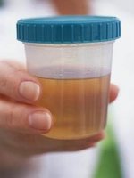
Appearance: Clear to deep amber (due to urochrome).
Odor: Slightly aromatic; may change with diet or disease.
Specific Gravity: 1.005–1.030 (correlates with osmolarity).
pH: 4.5–8.2 (average 6.0).
Chemical Composition: 95% water, 5% solutes (urea, NaCl, KCl, creatinine, uric acid).
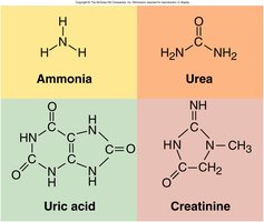
Nitrogenous Wastes
Urea: From amino acid catabolism (liver converts NH2 to urea).
Uric Acid: From nucleic acid breakdown.
Creatinine: From creatine phosphate metabolism.
Abnormal Constituents of Urine
Albuminuria: Presence of albumin (proteinuria).
Glucosuria: Presence of glucose (diabetes mellitus).
Hematuria: Presence of blood (UTI, trauma, stones).
Pyuria: Presence of pus (infection).
Ketonuria: Presence of ketones (starvation, diabetes).
Bilirubinuria: Presence of bilirubin (liver disease).
Urine Volume and Disorders
Normal and Abnormal Volumes
Normal: 1–2 L/day.
Polyuria: >2 L/day (diabetes, diuretics).
Oliguria: <500 mL/day (dehydration, kidney disease).
Anuria: <100 mL/day (renal failure).
Diabetes and Diuretics
Diabetes Mellitus: High blood glucose, glycosuria, polyuria.
Diabetes Insipidus: ADH deficiency, large urine volume.
Diuretics: Increase urine output, used for hypertension and heart failure (e.g., caffeine, alcohol).


Urinary Tract Anatomy
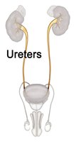
Ureters
Muscular tubes (~25 cm) transporting urine from renal pelvis to bladder via peristalsis.
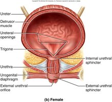
Urinary Bladder
Structure: Muscular sac with rugae for distension.
Trigone: Triangular area between ureter and urethra openings; common site for infection.
Capacity: 500 mL (moderately full), up to 800 mL (max).
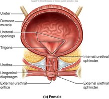
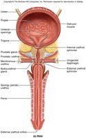
Urethra
Female: 3–4 cm (higher risk of UTI).
Male: 18 cm, passes through prostate and penis.
Sphincters: Internal (smooth muscle, involuntary) and external (skeletal muscle, voluntary).
Micturition (Voiding Urine)
Control of Urination
Bladder stretch receptors signal the micturition center when ~200 mL urine accumulates.
Results in bladder contraction and relaxation of internal sphincter; voluntary control via external sphincter.
Voluntary abdominal pressure can initiate urination if the bladder is not full.
Kidney Pathology
Common Disorders
Infection: Cystitis (bladder), pyelonephritis (kidney).
Trauma/Exercise: Can cause proteinuria or hematuria.
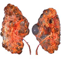
Polycystic Kidney Disease: Genetic disorder causing cysts, leading to kidney failure.
Kidney Stones (Renal Calculi): Hard deposits of minerals; cause pain and obstruction.
Renal Insufficiency: Inability to maintain homeostasis due to nephron loss.
Renal Cell Carcinoma: Cancer of kidney tubule cells; symptoms include pain, hematuria, and weight loss.
Hemodialysis
Artificial process to remove wastes from blood when kidneys fail.
Blood is filtered through a semipermeable membrane; typically performed several times per week.
Summary Table: Urinalysis Reference Values
Test | Normal Range | Abnormal Finding |
|---|---|---|
Specific Gravity | 1.005–1.030 | High/Low: dehydration/overhydration |
pH | 4.5–8.2 | Acidic/alkaline: diet, disease |
Protein | Negative | Proteinuria: kidney damage |
Glucose | Negative | Glucosuria: diabetes mellitus |
Blood | Negative | Hematuria: trauma, stones, infection |
Leukocytes | Negative | Pyuria: infection |
Ketones | Negative | Ketonuria: diabetes, starvation |
